. 2015 Jun;70(6):400-7.
doi: 10.6061/clinics/2015(06)03. Epub 2015 Jun 1.

Septic pulmonary embolism caused by a Klebsiella pneumoniae liver abscess: clinical characteristics, imaging findings, and clinical courses

Affiliations

Septic pulmonary embolism caused by a Klebsiella pneumoniae liver abscess: clinical characteristics, imaging findings, and clinical courses

Deng-Wei Chou et al. Clinics (Sao Paulo). 2015 Jun.

Abstract

Objectives: Septic pulmonary embolism caused by a Klebsiella (K.) pneumoniae liver abscess is rare but can cause considerable morbidity and mortality. However, clinical information regarding this condition is limited. This study was conducted to elucidate the full disease spectrum to improve its diagnosis and treatment.

Method: We reviewed the clinical characteristics, imaging findings, and clinical courses of 14 patients diagnosed with septic pulmonary embolism caused by a K. pneumoniae liver abscess over a period of 9 years.

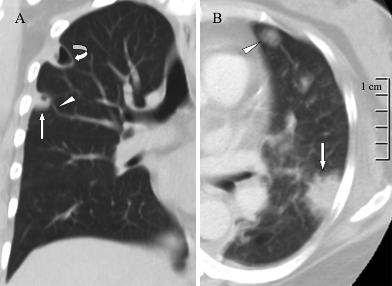
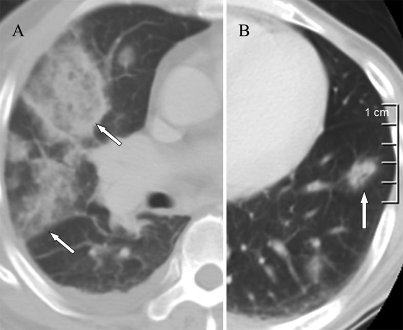
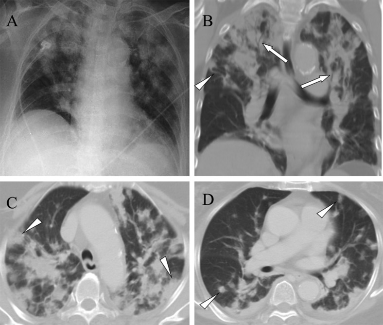
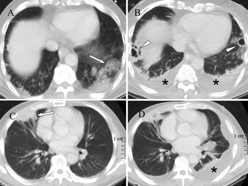
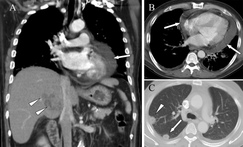
Results: The two most prevalent symptoms were fever and shortness of breath. Computed tomography findings included a feeding vessel sign (79%), nodules with or without cavities (79%), pleural effusions (71%), peripheral wedge-shaped opacities (64%), patchy ground-glass opacities (50%), air bronchograms within a nodule (36%), consolidations (21%), halo signs (14%), and lung abscesses (14%). Nine (64%) of the patients developed severe complications and required intensive care. According to follow-up chest radiography, the infiltrates and consolidations were resolved within two weeks, and the nodular opacities were resolved within one month. Two (14%) patients died of septic shock; one patient had metastatic meningitis, and the other had metastatic pericarditis.

Conclusion: The clinical presentations ranged from insidious illness with fever and respiratory symptoms to respiratory failure and septic shock. A broad spectrum of imaging findings, ranging from nodules to multiple consolidations, was detected. Septic pulmonary embolism caused by a K. pneumoniae liver abscess combined with the metastatic infection of other vital organs confers a poor prognosis.


Conflict of interest statement

No potential conflict of interest was reported.

Figures

Figure 1
Figure 1
A) A lung window of a coronal computed tomography scan shows a cavitary nodule (arrow) with a feeding vessel sign (arrowhead) and lung abscess (curved arrow) in the right upper lobe. B) A lung window of a cross-sectional computed tomography scan shows a peripheral wedge-shaped opacity (arrow) and ground-glass attenuation surrounding a pulmonary nodule (arrowhead) in the left upper lobe.
Figure 2
Figure 2
A) A lung window of a cross-sectional computed tomography scan shows multiple patchy ground-glass opacities (arrows) in the right lung. B) A lung window of a cross-sectional computed tomography scan shows air bronchograms within a nodule (arrow) in the left lung.
Figure 3
Figure 3
A) A chest radiograph reveals multiple alveolar consolidations, predominantly in the upper lung zones. B) A lung window of a coronal computed tomography scan shows multiple consolidations with air bronchograms (arrows) in the upper lobes. Additionally, a nodule with a feeding vessel sign (arrowhead) is shown. C) A lung window of a cross-sectional computed tomography scan shows multiple peripheral wedge-shaped (arrowheads) and D) nodular (arrowheads) opacities in the bilateral lungs.
Figure 4
Figure 4
A) A lung window of a cross-sectional computed tomography scan shows patchy ground-glass opacities in the left lower lobe (arrow). B) A repeat computed tomography scan obtained in the same image plane 7 days later shows new bilateral lung abscess (arrowheads) and pleural effusion (asterisks) formation. C) A lung window of a cross-sectional computed tomography scan shows a peripheral wedge-shaped opacity in the right middle lobe (arrow). D) A repeat computed tomography scan obtained in the same image plane 12 days later shows interval regression of the peripheral wedge-shaped opacity. A new left loculated pleural effusion (asterisk) formation is observed.
Figure 5
Figure 5
Pericardial effusion and septic pulmonary embolism caused by a Klebsiella pneumoniae liver abscess in a 73-year-old woman. A) A mediastinum window of a coronal computed tomography scan reveals a hypodense, hypovascular mass of approximately 5 cm in diameter in the S7 area of the right hepatic lobe (arrowheads) and fluid in the pericardial space (arrow). B) A mediastinum window of a cross-sectional computed tomography scan shows fluid in the pericardial space (arrows). C) A lung window of a cross-sectional computed tomography scan shows a peripheral wedge-shaped (arrowhead) and a peripheral nodular (arrow) opacity.

Similar articles

Cited by

References

    1. Bach AG, Restrepo CS, Abbas J, Villanueva A, Lorenzo Dus MJ, Schöpf R, et al. Imaging of nonthrombotic pulmonary embolism: biological materials, nonbiological materials, and foreign bodies. Eur J Radiol. 2013;82((3)):e120–41. doi: 10.1016/j.ejrad.2012.09.019. - DOI - PubMed
    1. Cook RJ, Ashton RW, Aughenbaugh GL, Ryu JH. Septic pulmonary embolism: presenting features and clinical course in 14 patients. Chest. 2005;128((1)):162–6. doi: 10.1378/chest.128.1.162. - DOI - PubMed
    1. Kuhlman JE, Fishman EK, Teigen C. Pulmonary septic emboli: diagnosis with CT. Radiology. 1990;174((1)):211–3. doi: 10.1148/radiology.174.1.2294550. - DOI - PubMed
    1. Huang RM, Naidich DP, Lubat E, Schinella R, Garay SM, McCauley DI. Septic pulmonary emboli: CT-radiographic correlation. AJR Am J Roentgenol. 1989;153((1)):41–5. doi: 10.2214/ajr.153.1.41. - DOI - PubMed
    1. Iwasaki Y, Nagata K, Nakanishi M, Natuhara A, Harada H, Kubota Y, et al. Spiral CT findings in septic pulmonary emboli. Eur J Radiol. 2001;37((3)):190–4. doi: 10.1016/S0720-048X(00)00254-0. - DOI - PubMed

MeSH terms