Review
. 2015 Jun;5(6):150062.
doi: 10.1098/rsob.150062.

LMO2 at 25 years: a paradigm of chromosomal translocation proteins

Affiliations
Review

LMO2 at 25 years: a paradigm of chromosomal translocation proteins

Jennifer Chambers et al. Open Biol. 2015 Jun.

Abstract

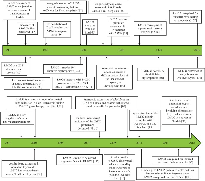
LMO2 was first discovered through proximity to frequently occurring chromosomal translocations in T cell acute lymphoblastic leukaemia (T-ALL). Subsequent studies on its role in tumours and in normal settings have highlighted LMO2 as an archetypical chromosomal translocation oncogene, activated by association with antigen receptor gene loci and a paradigm for translocation gene activation in T-ALL. The normal function of LMO2 in haematopoietic cell fate and angiogenesis suggests it is a master gene regulator exerting a dysfunctional control on differentiation following chromosomal translocations. Its importance in T cell neoplasia has been further emphasized by the recurrent findings of interstitial deletions of chromosome 11 near LMO2 and of LMO2 as a target of retroviral insertion gene activation during gene therapy trials for X chromosome-linked severe combined immuno-deficiency syndrome, both types of event leading to similar T cell leukaemia. The discovery of LMO2 in some B cell neoplasias and in some epithelial cancers suggests a more ubiquitous function as an oncogenic protein, and that the current development of novel inhibitors will be of great value in future cancer treatment. Further, the role of LMO2 in angiogenesis and in haematopoietic stem cells (HSCs) bodes well for targeting LMO2 in angiogenic disorders and in generating autologous induced HSCs for application in various clinical indications.

Keywords: LMO2; X chromosome-linked severe combined immuno-deficiency syndrome; angiogenesis; chromosomal translocations; haematopoiesis; leukaemia.


Figures

Figure 1.
Figure 1.
Milestones in LMO2 research: timeline indicating the major steps in LMO2 research from the gene discovery in 1990 to present.
Figure 2.
Figure 2.
Chromosomal translocations and X-SCID retroviral insertions associated with LMO2 gene activation. Diagram of the chromosomal bands of TCRA/D and TCRB and LMO2 involved in T cell ALL translocations resulting in LMO2 activation. Also indicated are the retroviral insertions found in the X-SCID gene therapy trial leukaemias (orange lines, with orientation of insertion indicated by orange arrows) [–11]. The distal, proximal [12] and intermediate [13] gene promoters are shown (black arrows). LMO2 comprises six exons (light green boxes, numbered) of which exons 4–6 (dark green boxes, numbered) are protein coding (green ribbon structure) drawn in USCF Chimaera [14] from PDB file 2XJY [15]. The coding region of LMO2 is unaltered after either the chromosomal translocations or the retroviral insertions. (Adapted from [16].)
Figure 3.
Figure 3.
Diagrammatic structure of the LIM-Only proteins and LMO2 amino acid sequence comparing human with mouse. (a) Schematic diagram of the LMO proteins showing the tandem arrangement of the two LIM-domains. Each domain comprises two zinc-finger-like structures, which coordinate a zinc atom between four residues. The two fingers of each domain are linked by two amino acid residues which are conserved between species and confer specificity of subsequent PPI. (b) An alignment of the human and mouse LMO2 proteins to illustrate species conservation and the homology and differences between them. Residues are highlighted to correlate with their position in the schematic structure shown in (a); green denotes cysteine, orange indicates histidine, yellow is aspartate while the key hinge region residue, phenylalanine 88, is highlighted in red [15].
Figure 4.
Figure 4.
Site-specific mechanism of LMO2 chromosomal translocations mediated by RAG recombinase. (a) TCR gene segments (V, D and J) are linked to RSS. Each segment has heptamer and nonamer sequences as indicated (and at the spacings in TCR V, D and J segments shown). (b) Heptamer sequences are found near LMO2 at chromosome 11p13 (green), specifically around translocation breakpoint regions denoted by black arrows [37]. Although not all were found to possess joining via the heptamer sequences, hallmark sequences of Rag recombinase were noted. The location of the proximal LMO2 promoter is shown by a red arrow. (c) Detailed analysis of the reciprocal translocation breakpoints of LMO2 has shown the involvement of the VDJ RAG recombinase in the translocations. An example of the organization of reciprocal t(7;11)(q35;p13) translocation regions is shown (lower part of the panel) compared with germline organization. Note the presence of a heptamer-like sequence at the junction of the 11p13 breakpoint [37].
Figure 5.
Figure 5.
Representation of the conformational flexibility of LMO2 in complex with LDB1 or inhibitors of LMO2. The pentameric LMO2-complex commonly found in erythroid lineage cells binds to a bipartite E-box-GATA motif [45] and is permitted or inhibited depending upon the stabilizing protein partner of LMO2 (a,d). LMO2 has been shown to nucleate variants of this complex, including the substitution of the GATA-factor and binding other bHLH transcription factor proteins [47], with resulting altered DNA recognition sequence binding specificity [48]. The structure and conformational stabilization of LMO2 (green) when bound with LDB1-LID (purple) [15] is indicated in (a). When LMO2 (shown in blue in panel (b)) is in complex with the anti-LMO2 VH (magenta), there is a conformational distortion, preventing nucleation of the LMO2-complex to the complex illustrated in (d) [49]. Superimposition of the normal LDB1- and VH-bound structures highlights the 23° contortion of the molecule when the anti-LMO2 VH is bound (e). The hinge region residue Phe88 (red), zinc atoms coordinated by the LIM-fingers (grey spheres) are indicated in both structures, drawn using UCSF Chimaera [14] from PDB accession codes 2XJY [15] and 4KFZ [49]. An in silico model of the structural effects on LMO2 induced by the peptide aptamer PA-207 is shown in (c). (Adapted from [50].)
Figure 6.
Figure 6.
Role of LMO2 in mouse development. Gene knock-out of mouse LMO2 has revealed its involvement in several aspects of haemaotopoiesis and blood vessel formation. LMO2 has a necessary role in primitive [24] (a) and definitive haematopoiesis [66] (b), and it is required for remodelling of the vascular networks as part of angiogenesis [67] (c). LMO2 has been shown to be expressed in the developing central nervous system [17] and tissue staining using lacZ knock-in mice showed specific regions of LMO2 expression [66] although no discernable knock-out brain phenotype has been noted to date.
Figure 7.
Figure 7.
Model of LMO2-mediated leukaemogenesis. Schematic diagram to represent the current understanding of the effect of LMO2 in LMO2-translocation positive T-ALL (a) and in the X-SCID gene therapy trial leukaemias (b) where LMO2 expression was activated through retroviral integration. (a) Fully mature CD4 or CD8 single positive (SP) T cells develop in the thymus through DN CD4/CD8 precursor populations, DN1–4, which are characterized by their progressive changing expression of CD25 and CD44 surface markers. The RAG recombinase genes are expressed from the DN2 stage onwards and promote TCR gene recombination events to produce functioning receptor molecules during the normal maturation cycle of the early thymoctyes. Although some cells progress through to mature CD4/CD8 double positive or SP T cells, thymus-specific transgenic Lmo2 mice exhibit a block in T cell differentiation with an accumulation of DN2–DN3 stage immature thymocytes (indicated), which after a long asymptomatic phase develop clonal T cell neoplasias [–89] after the acquisition of additional mutations [53]. This differentiation block coincides with the timing of RAG expression, directly mimicking the temporal occurrence, and effect of, chromosomal translocations involving the TCR genes and LMO2 loci [37]. Furthermore, the enforced expression of Lmo2 in the transgenics results in immature cells with acquiring self-renewal capacity, demonstrated by serial transplantation of the DN3 thymocytes [90]. (b) Following viral therapy in the X-SCID gene replacement trials, leukaemias were observed in which retroviral insertion had occurred into or upstream of the LMO2 gene and activated expression in thymocytes [–11]. Expanding from the LMO2 tumourigenesis model in (a), it would appear that leukaemogenesis in the X-SCID patients could arise from lymphoid progenitor cells with activated LMO2 expression migrating to the thymus, as part of the normal maturation process, and resulting in LMO2 conferring the DN2/3 differentiation block that was observed in the transgenic model, promoting self-renewal properties, accumulation of secondary mutations and finally the severe clinical adverse effects of leukaemias seen. (Adapted from [16].)

References

    1. Rabbitts TH. 1991. Translocations, master genes, and differences between the origins of acute and chronic leukemias. Cell 67, 641–644. (doi:10.1016/0092-8674(91)90057-6) - DOI - PubMed
    1. Rabbitts TH, Boehm T. 1991. Structural and functional chimerism results from chromosomal translocation in lymphoid tumors. Adv. Immunol. 50, 119–146. (doi:10.1016/s0065-2776(08)60824-x) - DOI - PubMed
    1. Lefranc MP, Rabbitts TH. 1985. Two tandemly organized human genes encoding the T-cell gamma constant-region sequences show multiple rearrangement in different T-cell types. Nature 316, 464–466. (doi:10.1038/316464a0) - DOI - PubMed
    1. Boehm T, Foroni L, Kaneko Y, Perutz MF, Rabbitts TH. 1991. The rhombotin family of cysteine-rich LIM-domain oncogenes: distinct members are involved in T-cell translocations to human chromosomes 11p15 and 11p13. Proc. Natl Acad. Sci. USA 88, 4367–4371. (doi:10.1073/pnas.88.10.4367) - DOI - PMC - PubMed
    1. Royer-Pokora B, Loos U, Ludwig WD. 1991. TTG-2, a new gene encoding a cysteine-rich protein with the LIM motif, is overexpressed in acute T-cell leukaemia with the t(11;14)(p13;q11). Oncogene 6, 1887–1893. - PubMed

Publication types

MeSH terms

Substances