. 2015 Jun 25:5:11585.
doi: 10.1038/srep11585.

Phosphorylation of Beet black scorch virus coat protein by PKA is required for assembly and stability of virus particles

Affiliations

Phosphorylation of Beet black scorch virus coat protein by PKA is required for assembly and stability of virus particles

Xiaofei Zhao et al. Sci Rep. .

Abstract

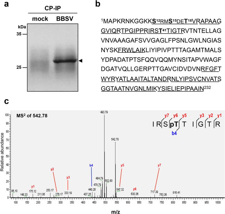
Plant virus coat proteins (CPs) play a fundamental role in protection of genomic RNAs, virion assembly, and viral movement. Although phosphorylation of several CPs during virus infection have been reported, little information is available about CP phosphorylation of the spherical RNA plant viruses. Here, we demonstrate that the CP of Beet black scorch virus (BBSV), a member of the genus Necrovirus, can be phosphorylated at threonine-41 (T41) by cAMP-dependent protein kinase (PKA)-like kinase in vivo and in vitro. Mutant viruses containing a T41A non-phosphorylatable alanine substitution, and a T41E glutamic acid substitution to mimic threonine phosphorylation were able to replicate but were unable to move systemically in Nicotiana benthamiana. Interestingly, the T41A and T41E mutants generated unstable 17 nm virus-like particles that failed to package viral genomic (g) RNA, compared with wild-type BBSV with 30 nm virions during viral infection in N. benthamiana. Further analyses showed that the T41 mutations had little effect on the gRNA-binding activity of the CP. Therefore, we propose a model whereby CP phosphorylation plays an essential role in long-distance movement of BBSV that involves formation of stable virions.


Conflict of interest statement

The authors declare no competing financial interests.

Figures

Figure 1
Figure 1. Identification of phosphorylation sites of CP by LC-MS/MS.
(a) Coomassie blue staining of CP immunoprecipitated (IP) from mock or BBSV inoculated N. benthamiana leaf tissues. The CP band (arrowhead) was digested in gel and then subjected to LC-MS/MS analysis. (b) Mass spectral sequence coverage (amino acids presented with underline) and identified phosphorylation sites (amino acids presented in bold) in the BBSV CP. (c) MS2 spectrum of the doubly charged phosphopeptide 38IRSpTTIGTR46 (m/z 542.78) containing the T41 phosphorylated residue. The colored highlights show b and y ions in the sequence and the corresponding peaks in the spectrum.
Figure 2
Figure 2. Accumulation of BBSVwt and phosphorylation site mutant viruses in N. benthanmiana.
N. benthamiana leaves exhibiting local (a) and systemic (e) symptoms after inoculation with BBSVwt and BBSV mutants containing non-phosphorylatable CP substitutions. ELISA detection with CP-specific antibodies of leaf extracts was conducted on inoculated leaves at 14 dpi (b) and upper leaves at 21 dpi (f). CP titers calculated from A405 ELISA values are indicated on the Y-axis. Viral gRNA replication and CP expression were analyzed in inoculated and upper leaves by Northern blot (c and g) and Western blot (d and h), respectively. The number ratios of symptom induction among the total inoculated plants are showed at the bottom of panels a and e.
Figure 3
Figure 3. Phosphorylation of BBSV CP by PKA in vivo and in vitro.
(a) Detection of phosphorylation of CP by PKA in N. benthamiana. Total protein was extracted from BBSVwt-inoculated leaves, and subjected to Western blots using phospho-(Ser/Thr) PKA substrate antibodies (α-pS/T, top panel, lane 2) or CP-specific antibodies (α-CP, bottom panel, lane 2). Then total extracted proteins from BBSVwt and BBSVT41A inoculated leaves were also immunoprecipitated with CP-specific antibodies and the same volumes of recovered CPwt and CPT41A were analyzed with CP antibodies (bottom panel, lanes 4 and 5). For the phospho-(Ser/Thr) PKA antibody tests, gels were loaded with three times the amounts used for the CP antibody analyses (top panel, lanes 4 and 5). The mobility of the CP band is indicated by arrowheads, and mock-inoculated N. benthamiana extracts served as negative controls (lanes 1 and 3). (b) Coomassie blue staining of His-tagged recombined CP (rCP, arrowhead) from uninduced (lane 1) or IPTG induced (lane 2) E. coli. The insoluble (lane 3) and soluble (lane 4) rCPs were separated by centrifugation and the soluble fraction was purified by Ni-NTA affinity chromatography (lanes 4–8), and analyzed by SDS-PAGE. Molecular size markers are indicated on the left and the rCP is indicated on the right by an arrowhead. (c) In vitro phosphorylation of rCP by purified CKII (lane 3), commercial CaMKII (lane 6) and commercial PKA (lane 9, arrowhead). Reactions containing GST-tagged kinase specific substrates served as positive controls (lanes 4, 7, and 10), and reactions lacking rCP (lanes 2, 5, and 8) or specific kinases (lane 1) served as negative controls. The autoradiography (top), and Coomassie blue stained loading controls (bottom) are shown. (d) Validation of in vitro rCP phosphorylation with the H-89 PKA specific inhibitor. Samples were treated with buffers lacking (lane 3) or containing 1 μM (lane 4) and 10 μM (lane 5) of H-89. Autoradiography and equal loading are shown in the top and bottom panels, respectively. (e) In vitro phosphorylation assays indicated that T41 is a PKA substrate. His-tagged CPwt, CPT41A, CPT41E and CPR39A were phosphorylated in vitro by PKA and autoradiographed (top panel, arrowhead), and loading controls are indicated at the bottom panel.
Figure 4
Figure 4. Accumulation of BBSVwt and mutant viruses in N. benthamiana.
N. benthamiana leaves exhibiting local (a) and systemic (e) BBSVwt and mutant viruses symptoms at 14 and 21 dpi. CP accumulation was determined by ELISA (b and f) and Western blot (d and h). Replication of viral RNA in inoculated and upper leaves was examined by Northern blot (c and g). The infection rate is indicated at the bottom of panels a and e.
Figure 5
Figure 5. Detection of viral accumulation in N. benthamiana protoplasts transfected with BBSVwt and T41 mutant viruses.
Total RNA extracted from BBSVWT and T41 mutant inoculated protoplasts at 20 or 60 hpi was subjected to Northern blot with a DIG-labeled BBSV-specific probe (a and c. top panel). EtBr staining shows the rRNA loading controls (a and c, bottom panel) and gRNA and sgRNAs are indicated on the left. Total proteins extracted from the protoplasts were subjected to Western blot with CP-specific antibodies (b and d, top panel). Equal proteins loading is verified by Coomassie blue staining (b and d, bottom panel), and uninfected protoplasts (mock) served as a negative control.
Figure 6
Figure 6. Analysis of particles from N. benthamiana leaves inoculated with BBSVwt and mutant viruses.
(a) Transmission electron microscopy (TEM) of VLPs obtained by partial and refined purification procedures from N. benthamiana plants inoculated with BBSVwt, BBSVT41A, BBSVT41E, and BBSVR39A transcripts. Inoculated leaf tissue was harvested at 14 dpi, extracted and observed by TEM (top panel). The VLPs were further enriched by partial or refined purification procedures and detected by TEM (middle and bottom panel). Mock-inoculated leaf samples served as controls (A, F, and K). Bars = 100 nm. More than 50 particles in each sample were measured and their average diameters are shown under the top panel. (b) VLP CP detection by Western blot with the CP antibodies used 5 μl samples from partial (middle panel) and refined (bottom panel) purifications. Total protein extracted from inoculated leaf tissues served as controls (top panel). (c) EtBr detection of viral RNA from partially purified BBSVwt and mutant preparations (2 μg) after 1% agarose gel electrophoresis (lanes 3–6). BBSVwt virions from the refined purifications served as a positive control to identify the encapsidated RNA (lane 1). Mock-inoculated leaf extracts subjected to the partial purification served as a negative control (lane 2).
Figure 7
Figure 7. RNA stability in N. benthamiana extracts and RNA-binding affinities of wt and T41 mutants.
(a) RNase protection assay in extracts from N. benthamiana infected with BBSVwt and T41 mutants. Inoculated leaves were ground and incubated in PIPE buffer at 37 °C for 30 (lanes 2, 5, and 8) or 60 mins (lanes 3, 6, and 9) to permit endogenous RNase degradation of unprotected RNA. The total RNAs were subsequently extracted and analyzed by Northern blot with a BBSV-specific probe. Bands corresponding to gRNAs and sgRNAs are indicated on the left. Total RNA was extracted as an untreated control immediately after leaves were ground (lanes 1, 4, and 7). (b) RNA binding abilities of rCPwt (lane 1), rCPT41A (lane 2), and rCPT41E (lane 3) determined by North-western blot assays with an anti-digoxigenin antibody (α DIG, top panel). BSA served as a negative control (lane 4). The bottom panel shows Coomassie blue stained protein loading controls.

Similar articles

Cited by

References

    1. Cui X. An icosahedral virus found in sugar beet. J. Shihezi Agric. College 10, 73–78 (1988).
    1. Merhvar M. & Bragard C. Beet black scorch virus in Iran is more diverse than anywhere. Phytopathol. 99, S84–S85 (2009).
    1. Weiland J. J. et al. Characterization of a US Isolate of Beet black scorch virus. Phytopathol. 97, 1245–1254 (2007). - PubMed
    1. Weiland J. J., Larson R. L., Freeman T. P. & Edwards M. C. First report of Beet black scorch virus in the United States. Plant Dis. 90, 828–828 (2006). - PubMed
    1. Koenig R. & Valizadeh J. Molecular and serological characterization of an Iranian isolate of Beet black scorch virus. Arch. Virol. 153, 1397–1400 (2008). - PubMed

Publication types

MeSH terms