. 2015 Aug 15;26(16):2927-38.
doi: 10.1091/mbc.E15-03-0159. Epub 2015 Jun 24.

Nuclear-localized CTP:phosphocholine cytidylyltransferase α regulates phosphatidylcholine synthesis required for lipid droplet biogenesis

Affiliations

Nuclear-localized CTP:phosphocholine cytidylyltransferase α regulates phosphatidylcholine synthesis required for lipid droplet biogenesis

Adam J Aitchison et al. Mol Biol Cell. .

Abstract

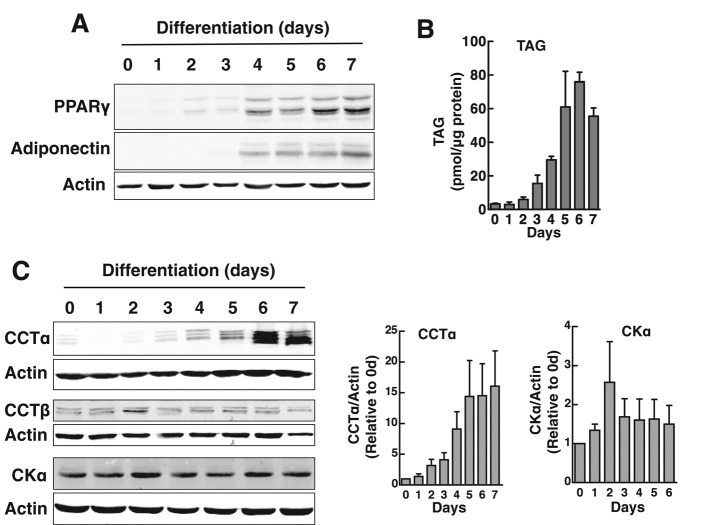
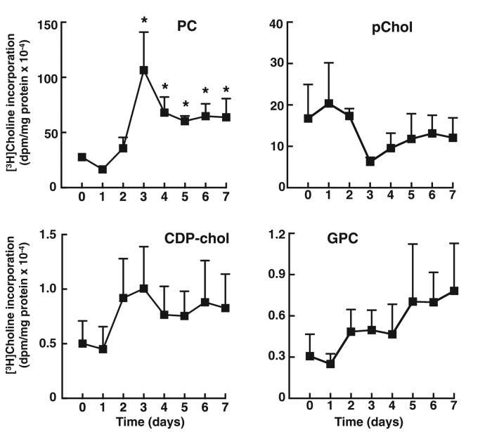
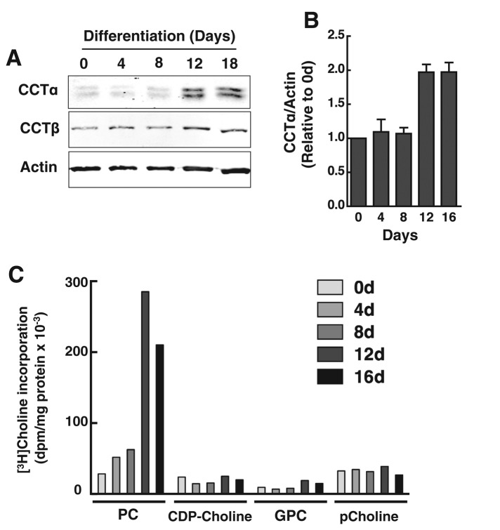
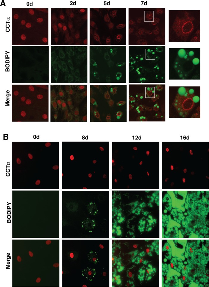
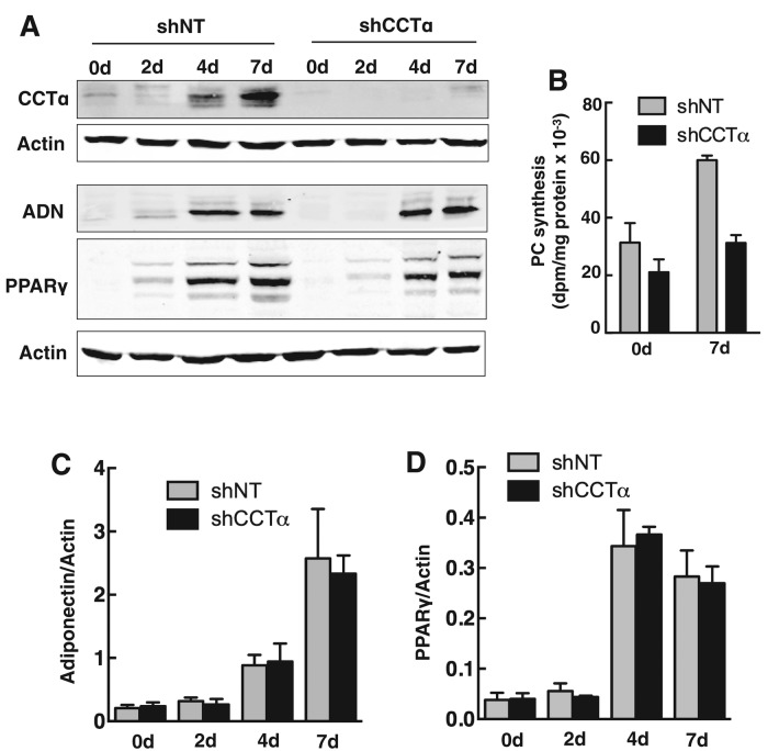
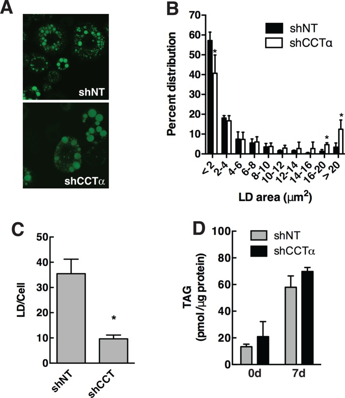
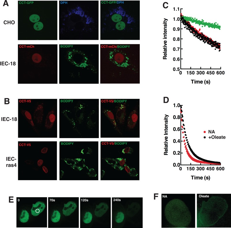
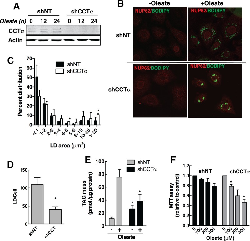
The reversible association of CTP:phosphocholine cytidylyltransferase α (CCTα) with membranes regulates the synthesis of phosphatidylcholine (PC) by the CDP-choline (Kennedy) pathway. Based on results with insect CCT homologues, translocation of nuclear CCTα onto cytoplasmic lipid droplets (LDs) is proposed to stimulate the synthesis of PC that is required for LD biogenesis and triacylglycerol (TAG) storage. We examined whether this regulatory mechanism applied to LD biogenesis in mammalian cells. During 3T3-L1 and human preadipocyte differentiation, CCTα expression and PC synthesis was induced. In 3T3-L1 cells, CCTα translocated from the nucleoplasm to the nuclear envelope and cytosol but did not associate with LDs. The enzyme also remained in the nucleus during human adipocyte differentiation. RNAi silencing in 3T3-L1 cells showed that CCTα regulated LD size but did not affect TAG storage or adipogenesis. LD biogenesis in nonadipocyte cell lines treated with oleate also promoted CCTα translocation to the nuclear envelope and/or cytoplasm but not LDs. In rat intestinal epithelial cells, CCTα silencing increased LD size, but LD number and TAG deposition were decreased due to oleate-induced cytotoxicity. We conclude that CCTα increases PC synthesis for LD biogenesis by translocation to the nuclear envelope and not cytoplasmic LDs.


Figures

FIGURE 1:
FIGURE 1:
CCTα expression is induced during differentiation of 3T3-L1 preadipocytes. 3T3-L1 cells were differentiated over a 7-d period as described in Materials and Methods. (A, B) Expression of adipocyte differentiation markers PPARγ and adiponectin was monitored by immunoblotting of total cell lysates. Accumulation of cellular TAG mass was assayed using a colorimetric assay. (C) Total cell lysates of differentiating 3T3-L1 cells were immunoblotted for CCTα, CCTβ, and choline kinase α (CKα). CCTα and CKα expression was quantified relative to actin using a Li-Cor (Lincoln, NE) imaging system and normalized to values at day 0. Results are the mean and SD of three or four separate experiments.
FIGURE 2:
FIGURE 2:
[3H]choline incorporation into PC is stimulated during differentiation of 3T3-L1 preadipocytes. 3T3-L1 cells were differentiated over a 7-d period as described in Materials and Methods. At the indicated times, cells were pulse labeled with [3H]choline (2 μCi/ml) for 3 h, and isotope incorporation into PC, phosphocholine (pChol), CDP-choline, and glycerophosphocholine (GPC) was measured. Results are the mean and SD of three experiments. *p < 0.05 compared to time 0 control.
FIGURE 3:
FIGURE 3:
Induction of CCTα and PC synthesis during human preadipocyte differentiation. (A, B) At the indicated times during a 16-d differentiation period, total cell lysates of primary human adipocytes were collected and immunoblotted for CCTα and CCTβ. CCTα expression was quantified relative to actin using a Li-Cor imaging system and normalized to day 0. Results are the mean and SD of three separate experiments. (C) At the indicated times during differentiation, primary human adipocytes were pulse labeled with [3H]choline (2 μCi/ml) for 3 h, and isotope incorporation into PC and water-soluble metabolites was measured. Results are the average of duplicate determinations from a representative experiment.
FIGURE 4:
FIGURE 4:
Nuclear CCTα does not associate with lipid droplets during 3T3-L1 or human preadipocyte differentiation. (A) At the indicated times during differentiation, 3T3-L1 cells were fixed, permeabilized, and immunostained with a CCTα primary antibody and an Alexa Fluor 594 secondary antibody. Lipid droplets were visualized with BODIPY-493/503. Right, enlarged images taken from the boxed areas for 7-d-differentiated cells. Images are 1-μm sections captured using a Zeiss LSM 510 META confocal microscope. Microscope settings for BODIPY imaging of 3T3-L1 cells were identical, but gain and pinhole settings for the 594-nm scan were adjusted due to increased CCTα overexpression at 5 and 7 d (Figure 1C). (B) Human preadipocytes differentiated over a 16-d period were fixed and immunostained for CCTα, and LDs were visualized with BODIPY-493/503 as described in A. Images are representative of three separate experiments.
FIGURE 5:
FIGURE 5:
CCTα silencing does not affect the expression of 3T3-L1 cell differentiation markers. (A) 3T3-L1 preadipocytes were stably transduced with lentivirus encoding shCCT or shNT as described in Materials and Methods. CCTα knockdown and shNT control cells were incubated in differentiation medium, and at the indicated times total cell lysates were immunoblotted for CCTα, adiponectin (ADN), and PPARγ. (B) Undifferentiated (0 d) or 7-d-differentiated (7 d) 3T3-L1 cells expressing shNT or shCCTα were pulse labeled for 3 h with [3H]choline (2 μCi/ml) to measure PC synthesis. Results are the mean and SD for three experiments. (C, D) Adiponectin and PPARγ expression in control shNT– and shCCTα-expressing 3T3-L1 cells during a 7-d differentiation period (shown in A) was quantified using a Li-Cor imaging system and normalized to actin expression. Results are the mean and SD of three separate experiments.
FIGURE 6:
FIGURE 6:
PC synthesis by the CDP-choline pathway regulates LD size in 3T3-L1 cells. (A) 3T3-L1 cells expressing shNT or shCCTα were either untreated or differentiated for 7 d, and LDs were visualized with BODIPY-493/503. (B) The size distribution of BODIPY-493/503 stained LDs in 7-d-differentiated control and knockdown cells was quantified using particle analysis features of ImageJ (version 1.48) and binned into area ranges. Results are the mean and SD of three or four fields of cells from three separate experiments. *p < 0.05 compared with matched shNT controls. (C) The total number of LDs in each cell was quantified from data in B. *p < 0.05 compared with matched shNT controls. (D) TAG mass was quantified in control and undifferentiated cells using an enzymatic assay. Results are the mean and SD of three experiments.
FIGURE 7:
FIGURE 7:
Localization of epitope-tagged CCTα in oleate-treated cells. (A) CHO and IEC-18 cells transiently expressing CCT-GFP or CCT-mCherry (CCT-mCh), respectively, were incubated with 400 μM oleate/BSA for 24 h. LDs were visualized by incubation with DPH or BODIPY-493/503. Spinning disk confocal images (1-μm sections) of live cells were captured as described in Materials and Methods. (B) IEC-18 and IEC-ras4 transiently expressing V5-tagged CCTα were treated with 400 μM oleate/BSA for 12 h before fixing and immunostaining with a V5-monoclonal and Alexa Fluor 594–conjugated secondary antibody. Images are 1-μm confocal sections captured using a Zeiss LSM510 META confocal microscope. (C) FLIP experiments in which the cytoplasm of CHO cells was repeatedly photobleached and relative fluorescence intensity of nuclear regions of interest (ROIs) were quantified. Green symbol, nonbleached cell; red symbol, no addition (NA); black symbol, oleate treated, 24 h. (D) FLIP experiment in which CHO cell nucleoplasm was repeatedly photobleached and the relative fluorescence intensity measured in a nuclear ROI. Results in C and D are the mean of four to nine cells from two separate experiments (error bars are removed for clarity). (E) FLIP analysis of an oleate-treated CHO cell (white circle indicates the photobleach area). (F) Nuclei of a control (NA) and oleate-treated CHO cell after the 10-min FLIP analysis shown in D. Image contrast and intensity was increased to visualize nuclear CCT-GFP.
FIGURE 8:
FIGURE 8:
PC synthesis by the CDP-choline pathway regulates LD size and TAG mass in oleate-treated IEC-18 cells. (A) IEC-18 cells were stably transduced with lentivirus encoding shNT or shCCTα and treated with or without 400 μM oleate/BSA for up to 24 h. Cell lysates were immunoblotted for CCTα to confirm silencing. (B) shNT- and shCCTα-transduced IEC-18 cells treated with or without 400 μM oleate/BSA for 24 h were immunostained with the nuclear pore protein NUP62 antibody and an Alexa Fluor 594 secondary antibody. LDs were visualized with BODIPY-493/503. (C, D) The size distribution and number of LDs in IEC-18 cells stimulated with oleate for 24 h was quantified as described in the legend to Figure 6. *p < 0.05 compared with matched shNT. (E) TAG mass was quantified in oleate-treated IEC-18 cells expressing shNT and shCCTα cells using a colorimetric assay. Results are the mean and SD of three experiments. (F) The viability of shNT- and shCCTα-transduced IEC-18 treated with 0–400 μM oleate was measured using an MTT assay. Results are expressed relative to untreated cells and are the mean and SD of three experiments. *p < 0.05 compared with similarly treated shNT cells.

References

    1. Agassandian M, Chen BB, Pulijala R, Kaercher L, Glasser JR, Mallampalli RK. Calcium-calmodulin kinase I cooperatively regulates nucleocytoplasmic shuttling of CCTalpha by accessing a nuclear export signal. Mol Biol Cell. 2012;23:2755–2769. - PMC - PubMed
    1. Agassandian M, Chen BB, Schuster CC, Houtman JC, Mallampalli RK. 14-3-3zeta escorts CCTalpha for calcium-activated nuclear import in lung epithelia. FASEB J. 2010;24:1271–1283. - PMC - PubMed
    1. Arnold RS, Cornell RB. Lipid regulation of CTP: phosphocholine cytidylyltransferase: electrostatic, hydrophobic, and synergistic interactions of anionic phospholipids and diacylglycerol. Biochemistry. 1996;35:9917–9924. - PubMed
    1. Arsenault DJ, Yoo BH, Rosen KV, Ridgway ND. ras-Induced up-regulation of CTP:phosphocholine cytidylyltransferase alpha contributes to malignant transformation of intestinal epithelial cells. J Biol Chem. 2013;288:633–643. - PMC - PubMed
    1. Bartz R, Li WH, Venables B, Zehmer JK, Roth MR, Welti R, Anderson RG, Liu P, Chapman KD. Lipidomics reveals that adiposomes store ether lipids and mediate phospholipid traffic. J Lipid Res. 2007;48:837–847. - PubMed

Publication types