Review
. 2015 Jul;18(7):942-52.
doi: 10.1038/nn.4043.

Diversity of astrocyte functions and phenotypes in neural circuits

Affiliations
Review

Diversity of astrocyte functions and phenotypes in neural circuits

Baljit S Khakh et al. Nat Neurosci. 2015 Jul.

Abstract

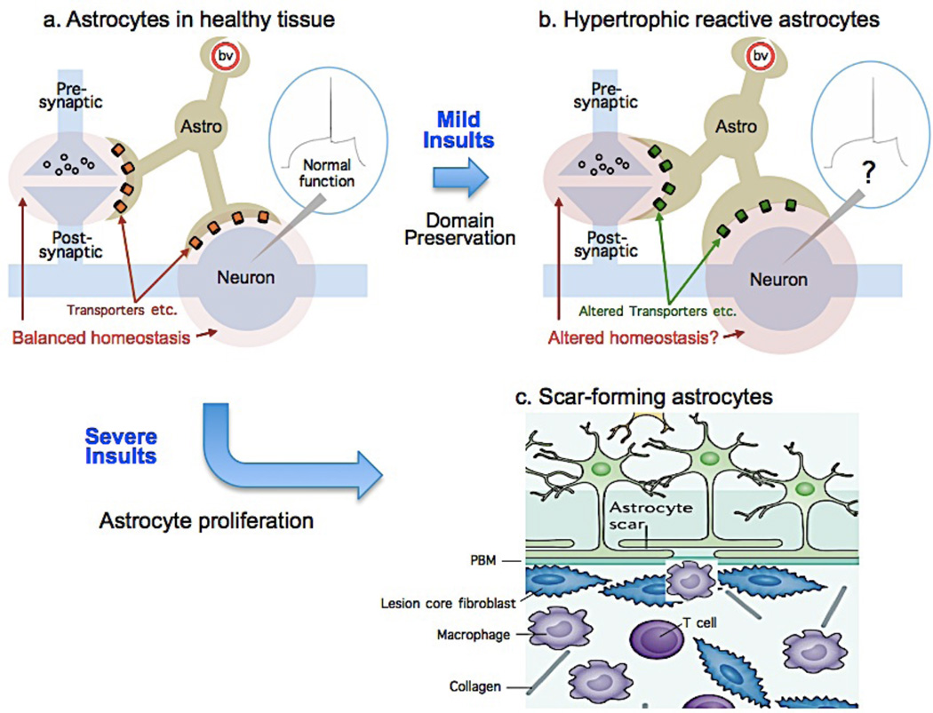
Astrocytes tile the entire CNS. They are vital for neural circuit function, but have traditionally been viewed as simple, homogenous cells that serve the same essential supportive roles everywhere. Here, we summarize breakthroughs that instead indicate that astrocytes represent a population of complex and functionally diverse cells. Physiological diversity of astrocytes is apparent between different brain circuits and microcircuits, and individual astrocytes display diverse signaling in subcellular compartments. With respect to injury and disease, astrocytes undergo diverse phenotypic changes that may be protective or causative with regard to pathology in a context-dependent manner. These new insights herald the concept that astrocytes represent a diverse population of genetically tractable cells that mediate neural circuit-specific roles in health and disease.


Figures

Figure 1
Figure 1. Astrocytes are morphologically complex cells
The images show hippocampal CA1 molecular layer protoplasmic astrocytes loaded with a fluorescent dye (Lucifer yellow). The images are from the Cell Centered Database, , at the National Center for Microscopy and Imaging Research (http://ccdb.ucsd.edu/index.shtm) and have Accession #1066, 1063. One can observe a soma, several major branches and thousands of branchlets and leaflets, which are discussed in the text
Figure 2
Figure 2. Morphologically complex astrocytes display well defined territories
The left hand image shows four hippocampal CA1 molecular layer astrocytes loaded with fluorescent dyes to illustrate how they do not encroach in to each other territory. This feature is easily apparent when examining how the astrocyte filled with a green dye does not markedly overlap with those filled with a red dye. The panel on the right shows a reconstruction of a Z-series of images and the yellow areas indicate the regions when the red and green signals overlap. The region of overlap is on the order of ~5%. These images are from the Cell Centered Database, , at the National Center for Microscopy and Imaging Research (http://ccdb.ucsd.edu/index.shtm) and have Accession #28.
Figure 3
Figure 3. Single astrocytes display diverse Ca2+ signals within territories
The image shows a CA1 region hippocampal astrocyte expressing cytosolic GCaMP6f. The colors indicate regions of interest that demarcate the soma (green), microdomains (yellow) and waves (red). The intensities of these regions are plotted as a function of time in the color coded traces (microdomains are shows in black for clarity). One can see that most Ca2+ signals occur in regions corresponding to branches and branchlets. The blue region demarcates the approximate territory, but such regions were not studied in detail. The figure and traces are reproduced from a published study.
Figure 4
Figure 4. Astrocytes in vivo respond with global Ca2+ elevations during electrical stimulation of the locus coeruleus, during locomotion and during startle responses
The lower cartoon illustrates the important experiment by Bekar and colleagues: they electrically stimulated LC neurons while monitoring Ca2+ signals in cortical astrocytes. They discovered global elevations in Ca2+ upon LC stimulation, which is illustrated in the upper cartoons. Similar global changes are seen when the mice are forced to walk or when they are startled, . Hence, during such responses, which may indicate a brain state change, the mode astrocyte Ca2+ signaling shifts from random Ca2+ microdomains and waves to global Ca2+ elevations. These global changes are mediated by noradrenaline release from LC projections, as discussed in the text.
Figure 5
Figure 5. Diverse astrocyte functional responses to tissue injury and disease
Cartoons illustrate astrocytes in healthy tissue (a) and two different functional changes in response to mild or severe insults. b: Mild insults trigger hypertrophic reactive astrogliosis in which astrocytes remain in situ, retain their domains and interactions with functioning neural cells but hypertrophy and undergo molecular and functional changes the consequences of which are not well defined. c: Severe insults that cause overt tissue damage trigger astrocyte proliferation from multiple potential sources. Newly proliferated scar-forming astrocytes are motile, intertwine their processes, interact primarily with fibrotic and inflammatory cells, and form borders around damaged and inflamed tissue. PBM, parenchymal basement membrane.

References

    1. Allen NJ, Barres BA. Neuroscience: Glia - more than just brain glue. Nature. 2009;457:675–677. - PubMed
    1. Herculano-Houzel S. The glia/neuron ratio: how it varies uniformly across brain structures and species and what that means for brain physiology and evolution. Glia. 2014;62:1377–1391. - PubMed
    1. Insel TR, Landis SC, Collins FS. Research priorities. The NIH BRAIN Initiative. Science. 2013;340:687–688. - PMC - PubMed
    1. De Felipe J. Cajal's butterflies of the soul: Science and art. Oxford University Press. 2010
    1. Oberheim NA, et al. Uniquely hominid features of adult human astrocytes. J Neurosci. 2009;29:3276–3287. - PMC - PubMed

Publication types

MeSH terms