. 2015 Jun 15;29(12):1285-97.
doi: 10.1101/gad.264234.115.

Homeostatic control of Hippo signaling activity revealed by an endogenous activating mutation in YAP

Affiliations

Homeostatic control of Hippo signaling activity revealed by an endogenous activating mutation in YAP

Qian Chen et al. Genes Dev. .

Abstract

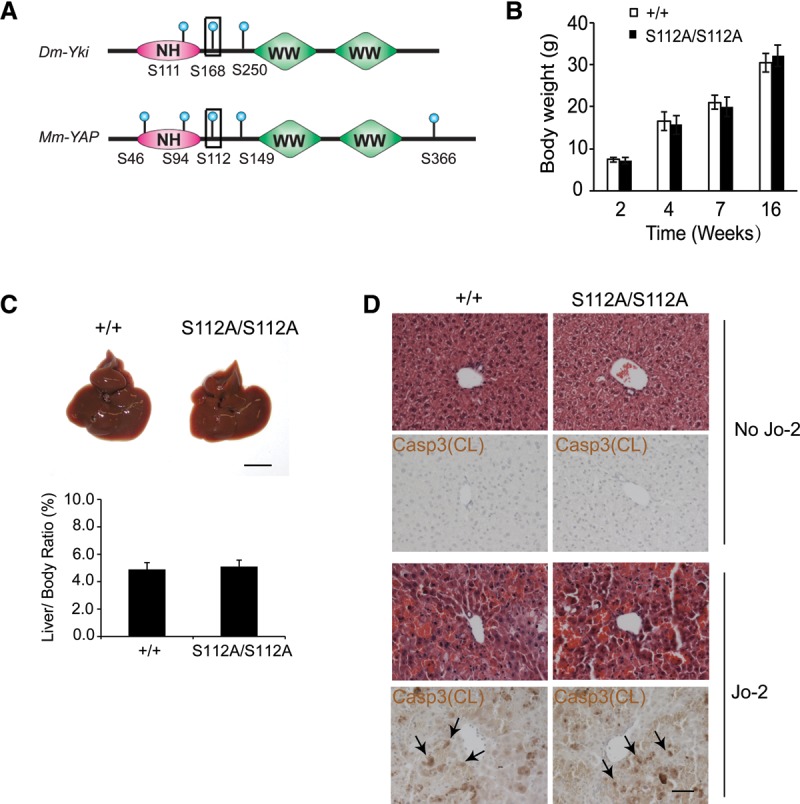
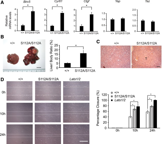
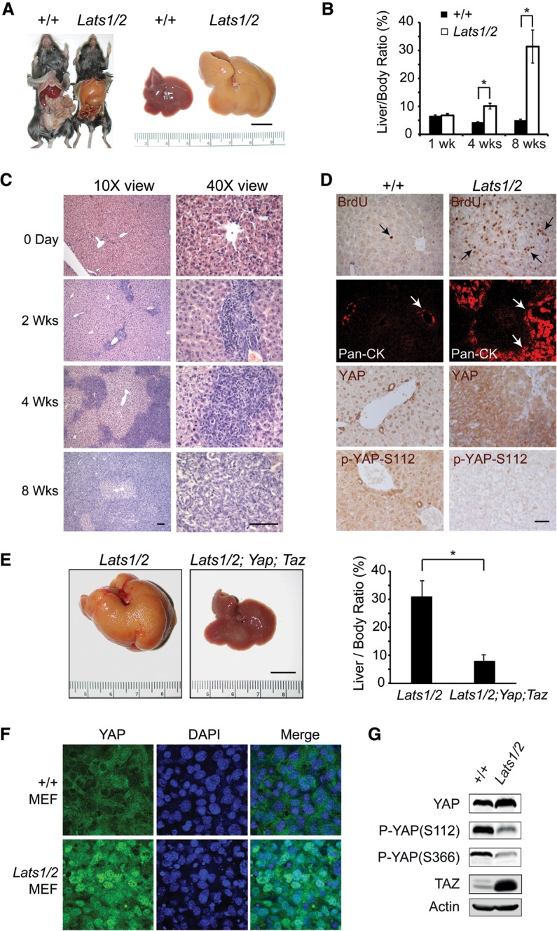
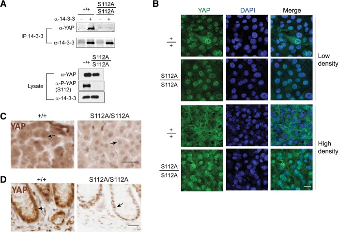
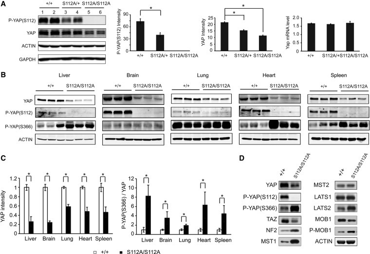
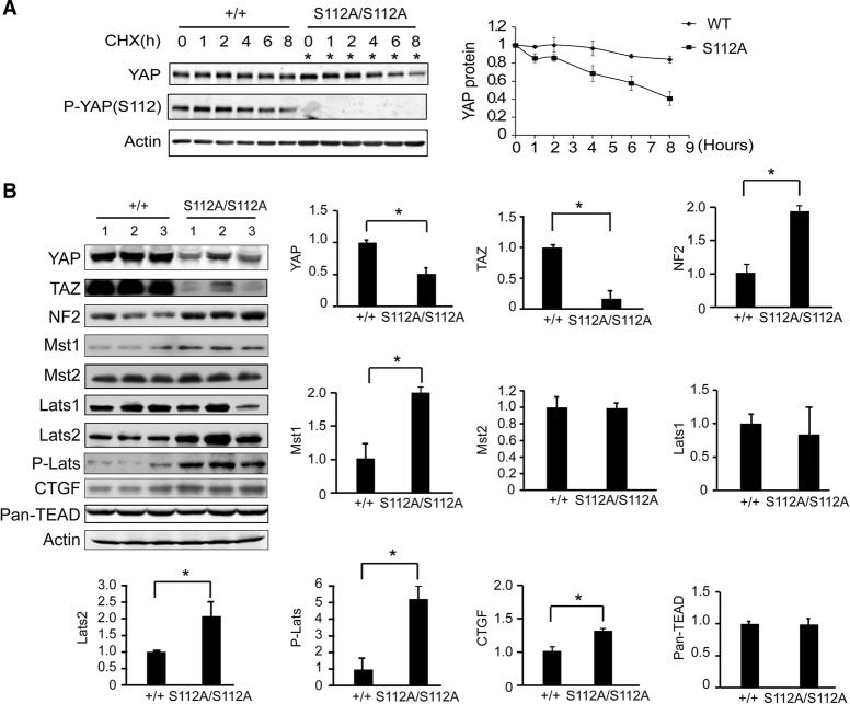
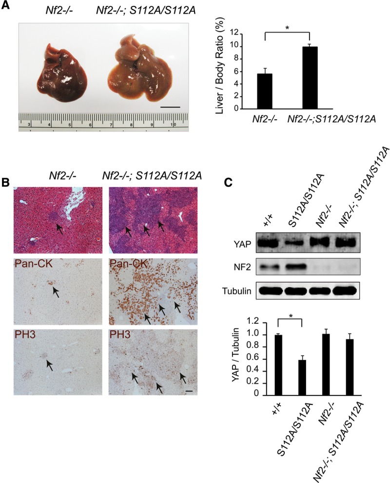
The Hippo signaling pathway converges on YAP to regulate growth, differentiation, and regeneration. Previous studies with overexpressed proteins have shown that YAP is phosphorylated by its upstream kinase, Lats1/2, on multiple sites, including an evolutionarily conserved 14-3-3-binding site whose phosphorylation is believed to inhibit YAP by excluding it from the nucleus. Indeed, nuclear localization of YAP or decreased YAP phosphorylation at this site (S168 in Drosophila, S127 in humans, and S112 in mice) is widely used in current literature as a surrogate of YAP activation even though the physiological importance of this phosphorylation event in regulating endogenous YAP activity has not been defined. Here we address this question by introducing a Yap(S112A) knock-in mutation in the endogenous Yap locus. The Yap(S112A) mice are surprisingly normal despite nuclear localization of the mutant YAP protein in vivo and profound defects in cytoplasmic translocation in vitro. Interestingly, the mutant Yap(S112A) mice show a compensatory decrease in YAP protein levels due to increased phosphorylation at a mammalian-specific phosphodegron site on YAP. These findings reveal a robust homeostatic mechanism that maintains physiological levels of YAP activity and caution against the assumptive use of YAP localization alone as a surrogate of YAP activity.

Keywords: 14-3-3; Hippo signaling; YAP oncoprotein; phosphorylation; tumorigenesis.


Figures

Figure 1.
Figure 1.
YAPS112 phosphorylation is dispensable for normal mouse development. (A) Schematic comparison of Drosophila Yki and murine YAP proteins showing the multiple HxRxxS phosphorylation sites, the WW domains, and the N-terminal homology (NH) domain required for Sd/TEAD binding. The conserved 14-3-3 site is also marked (boxed site). (B) Body weight of wild-type and YapS112A/S112A littermates. (C) Gross appearance of livers and quantification of the liver/body weight ratio from wild-type and YapS112A/S112A littermates at 2 mo of age. Data are mean ± SD from five animals of each genotype. Bar, 1 cm. (D) H&E and cleaved Caspase-3 staining of liver sections from 3-mo-old wild-type and YapS112A/S112A littermates before and 3 h after Jo-2 injection. Bar, 50 µm.
Figure 2.
Figure 2.
Mild gain of function of YAP in the YapS112A mice as revealed by YAP target expression and susceptibility to hepatocellular carcinogenesis. (A) Real-time PCR analysis of Birc5, Cyr61, Ctgf, Yap, and Taz mRNA levels in liver tissues of wild-type and YapS112A mice. Data are mean ± SD. n = 3 for each genotype. (*) P < 0.05, t-test. (B) Gross appearance of livers and quantification of liver/body weight ratio of wild-type and YapS112A mice 6.5 mo after diethylnitrosoamine (DEN) treatment. Data are mean ± SD from five animals of each genotype. (*) P < 0.05, t-test. Bar, 1 cm. (C) H&E staining of liver sections from wild-type and YapS112A mice 6.5 mo after DEN treatment. A large tumor in the YapS112A liver is indicated by “T.” Bar, 100 μm. (D) Wild-type, YapS112A/S112A, and Lats1/2 knockout mouse embryonic fibroblasts (MEFs) were subjected to wound healing assay. Shown are representative images of wound healing from 0 to 24 h after wound scratch. The white dashed lines mark the edges of the wound. Bar, 500 μm. Cell migration into the wound scratch was quantified as the percent wound closure relative to the open wound and compared with that of wild-type cells at each time point. All values are the means of triplicate experiments ± SD. (*) P < 0.05, t-test.
Figure 3.
Figure 3.
Lats1 and Lats2 are required for liver homeostasis by restricting YAP activity. (A) Gross appearance of control and Lats1−/−; Lats2flox/flox mouse livers 8 wk after Ad-Cre injection. Bar, 1 cm. (B) Quantification of liver/body ratio in wild-type and Lats1−/−; Lats2flox/flox littermates after Ad-Cre injection. Data are mean ± SD from five animals of each genotype. (*) P < 0.05, t-test. (C) H&E staining of liver sections from Lats1−/−; Lats2flox/flox animals before and after Ad-Cre injection. Bar, 50 μm. (D) BrdU, wide-spectrum CK (pan-CK), YAP, and phospho-YAP (S112) staining of liver sections from wild-type and Lats1−/−; Lats2flox/flox littermates 4 wk after Ad-Cre injection. Bar, 50 μm. (E) Gross appearance of livers and quantification of liver/body weight ratio of Lats1−/−; Lats2flox/flox and Lats1−/−; Lats2flox/flox; Yapflox/flox; Tazflox/flox mice 8 wk after Ad-Cre injection. Data are mean ± SD from five animals of each genotype. (*) P < 0.05, t-test. Bar, 1 cm. (F) Loss of cell contact-induced YAP translocation in the Lats1/2 mutant MEFs. Wild-type and Lats1/2 mutant MEFs grown at high density were immunostained for endogenous YAP (green) and nuclear dye DAPI (blue). Note the prominent nuclear staining of YAP in the Lats1/2 mutant MEFs compared with the wild-type MEFs. (G) Western blot analysis of cell lysates from the wild-type and Lats1/2 mutant MEFs.
Figure 4.
Figure 4.
S112 phosphorylation is required for 14-3-3 binding and cytoplasmic translocation of endogenous YAP induced by contact inhibition in cell culture or developmental Hippo signaling in intact tissues. (A) Coimmunoprecipitation assay. Cell lysates of wild-type and YapS112A/S112A MEFs were immunoprecipitated with α-14-3-3 antibody and immunoblotted with α-YAP antibody. (B) Loss of cell contact-induced YAP translocation in the YapS112A/S112A MEFs. Wild-type and YapS112A/S112A MEFs grown at low or high density were immunostained for endogenous YAP (green) and nuclear dye DAPI (blue). Endogenous YAP shows nuclear-to-cytoplasmic translocation at high cell density in the wild-type cells but not in the YapS112A/S112A cells. Bar, 50 μm. (C) Immunostaining of YAP in liver sections from wild-type and YapS112A mice. Note the more prominent nuclear localization of YAP in the YapS112A liver compared with the wild-type liver (arrows). Also note the overall decrease of YAP staining intensity in the YapS112A liver. Tissue sections were processed in parallel and stained under identical conditions. Bar, 50 μm. (D) Immunostaining of YAP in colon sections from wild-type and YapS112A mice. Note the decrease of overall YAP staining and the more prominent nuclear localization in the colonic epithelial cells in YapS112A mice compared with the wild-type mice (arrows). Tissue sections were processed and stained under identical conditions. Bar, 50 μm.
Figure 5.
Figure 5.
Compensatory reduction of YAP protein levels in the YapS112A mice. (A) Decreased YAP protein levels in the YapS112A livers. Liver lysates of wild-type, YapS112A/+, and YapS112A/S112A mice (4 mo old) were probed with the indicated antibodies. The graphs show quantification of YAP protein and mRNA levels. Note the absence of S112 phosphorylation and the decrease in YAP protein but not mRNA levels. Values are means ± SD. n = 3. (*) P < 0.05, t-test. (B) Decreased YAP protein levels and increased YAPS366 phosphorylation in multiple organs of the YapS112A mice (2 mo old). Note that although the p-YAPS366 level was comparable between mutant and wild-type brains, the ratio of p-YAPS366 relative to total YAP was higher in the mutant due to the dramatic decrease of YAP protein level (see quantification in C).Values are means ± SD. n = 3. (C) Quantification of relative YAP protein level (left graph) and P-YAP (S366)/YAP ratio (right graph) in different organs of wild-type and YapS112A mice. Data are mean ± SD from three animals of each genotype. (*) P < 0.05, t-test. (D) Western blot analysis of cell lysates from the wild-type and YapS112A MEFs.
Figure 6.
Figure 6.
Compensatory decrease of YAP stability and activation of the Hippo kinase cascade in the YapS112A mice. (A) Faster turnover of endogenous YAPS112A protein compared with endogenous wild-type (WT) YAP protein. Confluent cultures of wild-type and YapS112A MEFs were treated with 50 µg/mL CHX for different periods of time and probed with the indicated antibodies. A representative blot is shown. YAP protein levels were quantified by the LI-COR Odyssey imaging system from three parallel experiments, normalized to actin, and arbitrarily set as 1 at time 0 in the graph shown at the right. Note that twice as much YapS112A cell lysates were loaded in each lane to adjust for the lower levels of YAP protein in the YapS112A cells (*). (B) Western blot analysis of liver protein lysates from wild-type and YapS112A mice. The graphs show quantification of YAP, TAZ, NF2, Mst1, Mst2, Lats1, Lats2, p-Lats, TEAD1-4, and CTGF levels in mutant livers relative to wild-type livers. Data are mean ± SD from three animals for each genotype. (*) P < 0.05, t-test.
Figure 7.
Figure 7.
Feedback activation of Hippo signaling protects against developmental defects in the YapS112A animals. (A) Gross appearance of livers and quantification of liver/body weight ratio of Nf2flox/flox and Nf2flox/flox; YapS112A/S112A mice 8 wk after Ad-Cre injection. Data are mean ± SD from five animals of each genotype. (*) P < 0.05, t-test. Bar, 1 cm. (B) H&E, pan-CK, and phospho-Histone H3 (PH3) staining of liver sections from Nf2flox/flox and Nf2flox/flox; YapS112A/S112A littermates 8 wk after Ad-Cre injection. Note the increased BEC proliferation in Nf2flox/flox; YapS112A/S112A mutant livers compared with those of Nf2flox/flox mutant livers. Bar, 100 µm. (C) Western blot analysis of liver protein lysates from wild-type, YapS112A/S112A, Nf2flox/flox and Nf2flox/flox; YapS112A/S112A mice 2 mo after Ad-Cre injection. The graph shows quantification of YAP relative to tubulin from the four genotypes. Data are mean ± SD from three animals for each genotype. (*) P < 0.05, t-test.

References

    1. Barry ER, Camargo FD. 2013. The Hippo superhighway: signaling crossroads converging on the Hippo/Yap pathway in stem cells and development. Curr Opin Cell Biol 25: 247–253. - PubMed
    1. Benhamouche S, Curto M, Saotome I, Gladden AB, Liu CH, Giovannini M, McClatchey AI. 2010. Nf2/Merlin controls progenitor homeostasis and tumorigenesis in the liver. Genes Dev 24: 1718–1730. - PMC - PubMed
    1. Bossuyt W, Chen CL, Chen Q, Sudol M, McNeill H, Pan D, Kopp A, Halder G. 2014. An evolutionary shift in the regulation of the Hippo pathway between mice and flies. Oncogene 33: 1218–1228. - PMC - PubMed
    1. Cai J, Zhang N, Zheng Y, de Wilde RF, Maitra A, Pan D. 2010. The Hippo signaling pathway restricts the oncogenic potential of an intestinal regeneration program. Genes Dev 24: 2383–2388. - PMC - PubMed
    1. Camargo FD, Gokhale S, Johnnidis JB, Fu D, Bell GW, Jaenisch R, Brummelkamp TR. 2007. YAP1 increases organ size and expands undifferentiated progenitor cells. Curr Biol 17: 2054–2060. - PubMed

Publication types

MeSH terms