Review
. 2015 Jul 6;12(108):20150254.
doi: 10.1098/rsif.2015.0254.

Strategies for the chemical and biological functionalization of scaffolds for cardiac tissue engineering: a review

Review

Strategies for the chemical and biological functionalization of scaffolds for cardiac tissue engineering: a review

Marwa Tallawi et al. J R Soc Interface. .

Abstract

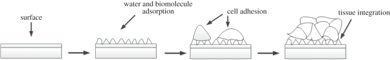
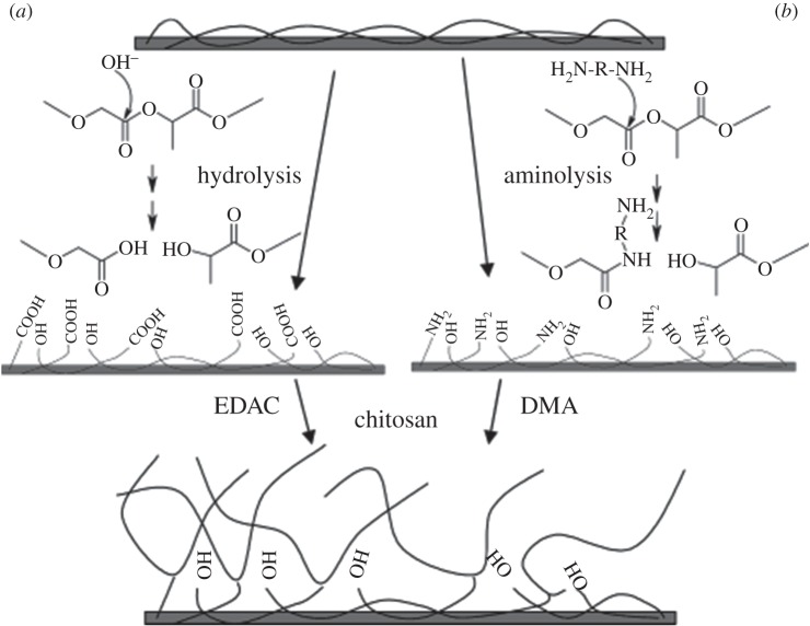
The development of biomaterials for cardiac tissue engineering (CTE) is challenging, primarily owing to the requirement of achieving a surface with favourable characteristics that enhances cell attachment and maturation. The biomaterial surface plays a crucial role as it forms the interface between the scaffold (or cardiac patch) and the cells. In the field of CTE, synthetic polymers (polyglycerol sebacate, polyethylene glycol, polyglycolic acid, poly-l-lactide, polyvinyl alcohol, polycaprolactone, polyurethanes and poly(N-isopropylacrylamide)) have been proven to exhibit suitable biodegradable and mechanical properties. Despite the fact that they show the required biocompatible behaviour, most synthetic polymers exhibit poor cell attachment capability. These synthetic polymers are mostly hydrophobic and lack cell recognition sites, limiting their application. Therefore, biofunctionalization of these biomaterials to enhance cell attachment and cell material interaction is being widely investigated. There are numerous approaches for functionalizing a material, which can be classified as mechanical, physical, chemical and biological. In this review, recent studies reported in the literature to functionalize scaffolds in the context of CTE, are discussed. Surface, morphological, chemical and biological modifications are introduced and the results of novel promising strategies and techniques are discussed.


Figures

Figure 1.
Figure 1.
The interaction of cells with biomaterials is governed by the surface properties of the biomaterial.
Figure 2.
Figure 2.
Schematic diagram showing three different surface chemical modification methods that can be exploited to modify the surface properties to add functional groups to the surface.
Figure 3.
Figure 3.
Schematic diagram showing two methods for the functionalization of PLGA surface, first activated (a) by hydrolysis (b) aminolysis and then further functionalized by covalently binding chitosan using EDAC or DMA, respectively. Reproduced with permission from ref. [156].
Figure 4.
Figure 4.
Chemical formula of maleimide investigated to attach heparin to PEG hydrogels [203].
Figure 5.
Figure 5.
Immobilization of bFGF and laminin on PLLA using di-NH2-PEG and heparin as linkers. Reproduced with permission from [178].
Figure 6.
Figure 6.
Simplified diagram showing the modulation of VEGF release profile acting on the carrier used to administer it (direct administration, encapsulation in nanoparticles, encapsulation in nanoparticles embedded in a scaffold). Reproduced with permission from [230]. (Online version in colour.)
Figure 7.
Figure 7.
Schematic representation of the scaffold fabrication process with dual GF delivery. Growth factors were incorporated into polymer scaffolds by either mixing with polymer particles before processing into scaffolds (VEGF), or pre-encapsulating the factor (PDGF) into polymer microspheres used to form scaffolds according to Richardson et al. Reproduced with permission from [249]. (Online version in colour.)

References

    1. Kajstura J, et al. 2010. Myocyte turnover in the aging human heart. Circ. Res. 107, 1374–1386. (10.1161/CIRCRESAHA.110.231498) - DOI - PubMed
    1. Improvement P, Goldberg LR. 2010. Heart failure. Ann. Intern. Med. 152, ITC61–15; quiz ITC616. - PubMed
    1. Birks E, Tansley P, Hardy J, George R, Bowles C, Burke M, Banner N, Khaghani A, Yacoub M. 2006. Left ventricular assist device and drug therapy for the reversal of heart failure. N. Engl. J. Med. 335, 1873–1884. (10.1056/NEJMoa053063) - DOI - PubMed
    1. Reffelmann T, Kloner RA. 2003. Cellular cardiomyoplasty—cardiomyocytes, skeletal myoblasts, or stem cells for regenerating myocardium and treatment of heart failure? Cardiovasc. Res. 58, 358–368. (10.1016/S0008-6363(02)00739-3) - DOI - PubMed
    1. Laflamme M, Murry C. 2005. Regenerating the heart. Nat. Biotechnol. 23, 845–856. (10.1038/nbt1117) - DOI - PubMed

Publication types

Substances