. 2015 Jun 24;35(25):9424-31.
doi: 10.1523/JNEUROSCI.0490-15.2015.

Role of Striatal Cholinergic Interneurons in Set-Shifting in the Rat

Affiliations

Role of Striatal Cholinergic Interneurons in Set-Shifting in the Rat

Sho Aoki et al. J Neurosci. .

Abstract

The ability to change strategies in different contexts is a form of behavioral flexibility that is crucial for adaptive behavior. The striatum has been shown to contribute to certain forms of behavioral flexibility such as reversal learning. Here we report on the contribution of striatal cholinergic interneurons-a key element in the striatal neuronal circuit-to strategy set-shifting in which an attentional shift from one stimulus dimension to another is required. We made lesions of rat cholinergic interneurons in dorsomedial or ventral striatum using a specific immunotoxin and investigated the effects on set-shifting paradigms and on reversal learning. In shifting to a set that required attention to a previously irrelevant cue, lesions of dorsomedial striatum significantly increased the number of perseverative errors. In this condition, the number of never-reinforced errors was significantly decreased in both types of lesions. When shifting to a set that required attention to a novel cue, rats with ventral striatum lesions made more perseverative errors. Neither lesion impaired learning of the initial response strategy nor a subsequent switch to a new strategy when response choice was indicated by a previously relevant cue. Reversal learning was not affected. These results suggest that in set-shifting the striatal cholinergic interneurons play a fundamental role, which is dissociable between dorsomedial and ventral striatum depending on behavioral context. We propose a common mechanism in which cholinergic interneurons inhibit neurons representing the old strategy and enhance plasticity underlying exploration of a new rule.

Keywords: behavioral flexibility; cholinergic interneuron; rat; set-shifting; striatum.


Figures

Figure 1.
Figure 1.
A flow chart of the behavioral paradigms. A yellow circle indicates a visual cue. In set-shifting, three different experimental conditions require animals to attend to a novel cue (A, Condition 1), to a previously relevant cue (B, Condition 2), or to a previously irrelevant cue (C, Condition 3).
Figure 2.
Figure 2.
Histological verification of specific lesions of cholinergic interneurons. A, Representative coronal sections of the rat striatum show intact NeuN staining but clear ablation of the cholinergic interneurons in ChAT staining in lesioned cases (DMS or VS). Scale bar, 1 mm. B, An example of a small nonspecific lesion indicated by a dashed area (NeuN) where there are sparse cells. C, D, The smallest (black) and largest cases (gray) of cholinergic interneuronal ablation in each group are drawn separately in three conditions. Distance from bregma is indicated to the left. The size of lesions in DMS (C) and VS (D) appears equivalent across conditions. LV, lateral ventricle; CC, corpus callosum; AC, anterior commissure.
Figure 3.
Figure 3.
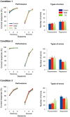
Behavioral performance and types of errors in strategy set-shifting. Percentage of correct responses in both response and visual cue strategy (left), types of errors committed over 10 sessions of visual cue strategy (middle), and early and late components of never-reinforced errors (right) are shown for each experimental condition. Set-shifting required attention to a novel stimulus (A), to a previously relevant stimulus (B), and to a previously irrelevant stimulus (C). Final group size is as follows: condition 1, n = 16 (control), n = 19 (DMS), n = 14 (VS); condition 2, n = 13 (control), n = 18 (DMS), n = 14 (VS); condition 3, n = 21 (control), n = 17 (DMS), n = 16 (VS). Data are shown as means ± SEM. *p < 0.05, **p < 0.01, and ***p < 0.001.
Figure 4.
Figure 4.
Behavioral performance and types of errors in retraining and reversal of the response strategy. Percentage of correct responses in retraining of the original response strategy and its reversal (left) and types of errors committed during reversal learning (right) are shown for each condition. Final group size is the same as Figure 3. Data are means ± SEM.

References

    1. Aosaki T, Graybiel AM, Kimura M. Effect of the nigrostriatal dopamine system on acquired neural responses in the striatum of behaving monkeys. Science. 1994;265:412–415. doi: 10.1126/science.8023166. - DOI - PubMed
    1. Aosaki T, Kimura M, Graybiel AM. Temporal and spatial characteristics of tonically active neurons of the primate's striatum. J Neurophysiol. 1995;73:1234–1252. - PubMed
    1. Apicella P. Leading tonically active neurons of the striatum from reward detection to context recognition. Trends Neurosci. 2007;30:299–306. doi: 10.1016/j.tins.2007.03.011. - DOI - PubMed
    1. Bradfield LA, Hart G, Balleine BW. The role of the anterior, mediodorsal, and parafascicular thalamus in instrumental conditioning. Front Syst Neurosci. 2013a;7:51. doi: 10.3389/fnsys.2013.00051. - DOI - PMC - PubMed
    1. Bradfield LA, Bertran-Gonzalez J, Chieng B, Balleine BW. The thalamostriatal pathway and cholinergic control of goal-directed action: interlacing new with existing learning in the striatum. Neuron. 2013b;79:153–166. doi: 10.1016/j.neuron.2013.04.039. - DOI - PMC - PubMed

Publication types

LinkOut - more resources