Clinical and imaging assessment of acute combat mild traumatic brain injury in Afghanistan
- PMID: 26109715
- PMCID: PMC4516289
- DOI: 10.1212/WNL.0000000000001758
Clinical and imaging assessment of acute combat mild traumatic brain injury in Afghanistan
Abstract
Objective: To evaluate whether diffusion tensor imaging (DTI) will noninvasively reveal white matter changes not present on conventional MRI in acute blast-related mild traumatic brain injury (mTBI) and to determine correlations with clinical measures and recovery.
Methods: Prospective observational study of 95 US military service members with mTBI enrolled within 7 days from injury in Afghanistan and 101 healthy controls. Assessments included Rivermead Post-Concussion Symptoms Questionnaire (RPCSQ), Post-Traumatic Stress Disorder Checklist Military (PCLM), Beck Depression Inventory (BDI), Balance Error Scoring System (BESS), Automated Neuropsychological Assessment Metrics (ANAM), conventional MRI, and DTI.
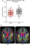
Results: Significantly greater impairment was observed in participants with mTBI vs controls: RPCSQ (19.7 ± 12.9 vs 3.6 ± 7.1, p < 0.001), PCLM (32 ± 13.2 vs 20.9 ± 7.1, p < 0.001), BDI (7.4 ± 6.8 vs 2.5 ± 4.9, p < 0.001), and BESS (18.2 ± 8.4 vs 15.1 ± 8.3, p = 0.01). The largest effect size in ANAM performance decline was in simple reaction time (mTBI 74.5 ± 148.4 vs control -11 ± 46.6 milliseconds, p < 0.001). Fractional anisotropy was significantly reduced in mTBI compared with controls in the right superior longitudinal fasciculus (0.393 ± 0.022 vs 0.405 ± 0.023, p < 0.001). No abnormalities were detected with conventional MRI. Time to return to duty correlated with RPCSQ (r = 0.53, p < 0.001), ANAM simple reaction time decline (r = 0.49, p < 0.0001), PCLM (r = 0.47, p < 0.0001), and BDI (r = 0.36 p = 0.0005).
Conclusions: Somatic, behavioral, and cognitive symptoms and performance deficits are substantially elevated in acute blast-related mTBI. Postconcussive symptoms and performance on measures of posttraumatic stress disorder, depression, and neurocognitive performance at initial presentation correlate with return-to-duty time. Although changes in fractional anisotropy are uncommon and subtle, DTI is more sensitive than conventional MRI in imaging white matter integrity in blast-related mTBI acutely.
© 2015 American Academy of Neurology.
Figures


Comment in
-
Comment: Does brain DTI MRI aid diagnosis of battlefield concussion?Neurology. 2015 Jul 21;85(3):226. doi: 10.1212/WNL.0000000000001771. Epub 2015 Jun 24. Neurology. 2015. PMID: 26109709 No abstract available.
References
-
- Hayward P. Traumatic brain injury: the signature of modern conflicts. Lancet Neurol 2008;7:200–201. - PubMed
-
- Warden D. Military TBI during the Iraq and Afghanistan wars. J Head Trauma Rehabil 2006;21:398–402. - PubMed
-
- Hoge CW, McGurk D, Thomas JL, Cox AL, Engel CC, Castro CA. Mild traumatic brain injury in U.S. Soldiers returning from Iraq. N Engl J Med 2008;358:453–463. - PubMed
-
- Practice parameter: the management of concussion in sports (summary statement). Report of the Quality Standards Subcommittee. Neurology 1997;48:581–585. - PubMed
-
- Cantu RC. Return to play guidelines after a head injury. Clin Sports Med 1998;17:45–60. - PubMed
Publication types
MeSH terms
LinkOut - more resources
Full Text Sources
Research Materials
