. 2015 Sep;89(18):9213-31.
doi: 10.1128/JVI.01213-15. Epub 2015 Jun 24.

Patient-Specific Neutralizing Antibody Responses to Herpes Simplex Virus Are Attributed to Epitopes on gD, gB, or Both and Can Be Type Specific

Affiliations

Patient-Specific Neutralizing Antibody Responses to Herpes Simplex Virus Are Attributed to Epitopes on gD, gB, or Both and Can Be Type Specific

Tina M Cairns et al. J Virol. 2015 Sep.

Abstract

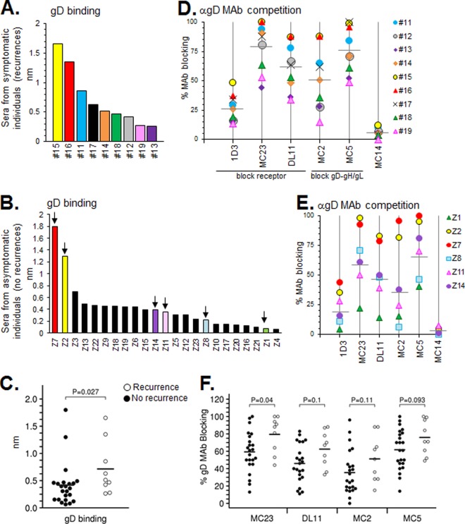
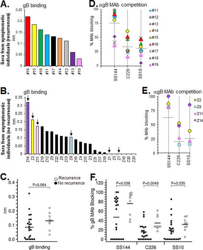
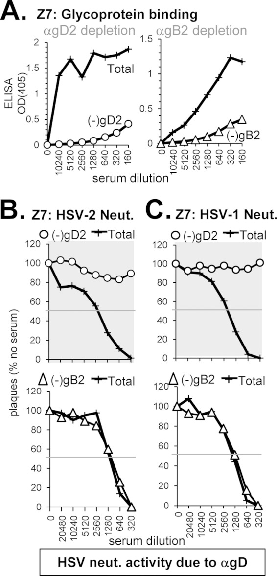
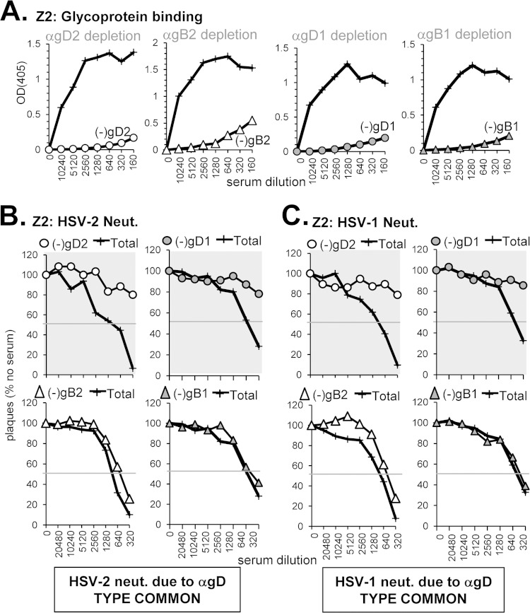
Herpes simplex virus 1 (HSV-1) and HSV-2 infect many humans and establish a latent infection in sensory ganglia. Although some infected people suffer periodic recurrences, others do not. Infected people mount both cell-mediated and humoral responses, including the production of virus-neutralizing antibodies (Abs) directed at viral entry glycoproteins. Previously, we examined IgGs from 10 HSV-seropositive individuals; all neutralized virus and were directed primarily against gD or gD+gB. Here, we expand our studies and examine 32 additional sera from HSV-infected individuals, 23 of whom had no recurrent disease. Using an Octet RED96 system, we screened all 32 serum samples directly for both glycoprotein binding and competition with known neutralizing anti-gD and -gB monoclonal Abs (MAbs). On average, the recurrent cohort exhibited higher binding to gD and gB and had higher neutralization titers. There were similar trends in the blocking of MAbs to critical gD and gB epitopes. When we depleted six sera of Abs to specific glycoproteins, we found different types of responses, but always directed primarily at gD and/or gB. Interestingly, in one dual-infected person, the neutralizing response to HSV-2 was due to gD2 and gB2, whereas HSV-1 neutralization was due to gD1 and gB1. In another case, virus neutralization was HSV-1 specific, with the Ab response directed entirely at gB1, despite this serum blocking type-common anti-gD and -gB neutralizing MAbs. These data are pertinent in the design of future HSV vaccines since they demonstrate the importance of both serotypes of gD and gB as immunogens.

Importance: We previously showed that people infected with HSV produce neutralizing Abs directed against gD or a combination of gD+gB (and in one case, gD+gB+gC, which was HSV-1 specific). In this more extensive study, we again found that gD or gD+gB can account for the virus neutralizing response and critical epitopes of one or both of these proteins are represented in sera of naturally infected humans. However, we also found that some individuals produced a strong response against gB alone. In addition, we identified type-specific contributions to HSV neutralization from both gD and gB. Contributions from the other entry glycoproteins, gC and gH/gL, were minimal and limited to HSV-1 neutralization. Knowing the variations in how humans see and mount a response to HSV will be important to vaccine development.


Figures

FIG 1
FIG 1
Virus neutralization. Sample sera were tested for their ability to neutralize HSV (types 1 and 2). Plaque numbers were determined for each sample and plotted as a percentage of plaques obtained in the absence of human serum (y axis). Serum dilutions are indicated on the x axis. Sera were obtained from either long-term-infected individuals with recurrent disease (symptomatic) (A) or individuals with no (zero) recurrence of disease (asymptomatic) (B). For samples from nonrecurrent individuals, a select set of sera are graphed in panel B. HSV neutralization data for all of the samples are provided in Table 2. (C) Statistical analysis of the neutralizing titer of patient sera from individuals with recurrent or nonrecurrent disease. The data from individuals with no clinical HSV recurrence are represented with black circles, individuals with HSV recurrence are represented with white circles, and horizontal black bars indicate the mean value. Symptomatic individuals produced a higher titer of neutralizing antibodies on average, but the variance between individuals resulted in a P value above 0.05.
FIG 2
FIG 2
Samples from HSV-infected humans, serum versus IgG. Antibody binding to gB was tested using purified IgG on a BIAcore 3000 biosensor (A) or using serum on an Octet RED96 system (B) as described in Materials and Methods. Relative binding units (RU) are indicated on the y axis. The averages of at least two experiments are shown; error bars indicate the standard errors. (C) Blocking of neutralizing anti-gB MAbs via human subject IgG (BIAcore) or sera (Octet). Binding curves for the association of test MAbs (SS144, C226, and SS10) to gB are shown. Black lines indicate MAb binding to gB that was not exposed to human subject samples (positive control). Gray dotted lines denote samples that block MAb binding less than 25% that of control. The curves reflect the extent to which Abs within the serum blocked MAb binding (steep curve, close to the black control curve = no MAb blocking; shallow curve = MAb blocking). The data using human IgG on the BIAcore for these six samples was first reported in Cairns et al. (35).
FIG 3
FIG 3
(A and B) Antibody binding to gD was tested using human sera on an Octet RED96 system as described in Materials and Methods. Samples are ranked from high to low for gD binding. (D and E) Blocking of neutralizing anti-gD MAbs via human subject serum. Each point indicates the percent blocking activity (compared to a no-Ab control) against the MAbs tested. Horizontal gray bars denote average blocking of that particular MAb among the samples. Sera from individuals with recurrent infection are shown in panels A and D, while those without are shown in panels B and E. For those with no recurrences, only selected samples from panel B (arrows) were graphed in panel E. Samples are color-coded the same between gD binding and MAb competition graphs. Anti-gD MAb competition data for all of the nonrecurrent samples are provided in Table 2. (C and F) Statistical analysis of the gD binding and MAb blocking capability of patient sera from individuals with recurrent or nonrecurrent disease. The data from individuals with no clinical HSV recurrence are represented with black circles, individuals with HSV recurrence are represented with white circles, and horizontal black bars indicate the mean value. Using Octet data from panels A and B, we found significantly more Abs targeted gD in recurring individuals than nonrecurring individuals (C). Likewise, individuals with recurrent disease produced more Abs directed toward all of the gD MAb epitopes we studied, and yet only the MC23 epitope reached statistical significance (F).
FIG 4
FIG 4
(A to F) Human sera binding to gB, competition with neutralizing anti-gB MAbs, and statistical analysis of these data was set up in the same manner as described for gD in Fig. 2. For sera from individuals with no recurrent disease, only selected samples from panel B (arrows) were graphed in panel E. Anti-gB MAb competition data for all of the nonrecurrent samples is provided in Table 2. Samples Z1 and Z7, which did not bind gB in panel B, were not tested for MAb competition in panel E. Those with recurrent infections produced Abs that exhibited greater gB binding and anti-gB MAb competition; blocking of all three anti-gB epitopes tested was statistically different between recurrent and nonrecurrent individuals.
FIG 5
FIG 5
Depletion of gD- and gB-specific Abs from human serum sample Z7. To deplete human serum of glycoprotein-specific Abs, serum was incubated with biotinylated gD2 or gB2 that was bound to streptavidin-coated magnetic beads. (A) After depletion, the sample supernatant was tested by ELISA for binding to soluble gD2 (○) or gB2 (△); the optical density at 405 nm (OD405) is shown on the y axis, and the serum dilution is indicated on the x axis of each ELISA graph. Next, the depleted sample was tested for the ability to neutralize either HSV-2 (B) or HSV-1 (C). Plaque numbers were determined for each sample and plotted as a percentage of plaques obtained in the absence of human serum (y axis). Serum dilutions are indicated on the x axis. Curves where the neutralization activity was successfully depleted are highlighted with a gray background.
FIG 6
FIG 6
Depletion of glycoprotein-specific Abs from human serum sample Z8. (A) Antibody depletion of serum was carried out as described in Fig. 4A. Depletion with gD1 is indicated with gray circles, and that of gB1 is indicated with gray triangles. The depleted serum was tested for neutralization activity against either HSV-2 (B) or HSV-1 (C) as in Fig. 4B and C, respectively. The bottom row in panel B and panel C depict samples where both Ab against gD and gB have been depleted (squares, white for type 2 and gray for type 1).
FIG 7
FIG 7
Depletion of glycoprotein-specific Abs from human serum sample Z2. (A) Antibody depletion of serum was carried out as described in Fig. 5A. The depleted serum was tested for neutralization activity against either HSV-2 (B) or HSV-1 (C) as in Fig. 5B and C, respectively.
FIG 8
FIG 8
Depletion of glycoprotein-specific Abs from human serum sample 16. (A) Antibody depletion of serum was carried out as described in Fig. 5A. The depleted serum was tested for neutralization activity against either HSV-2 (B) or HSV-1 (C) as in Fig. 5B and C, respectively.
FIG 9
FIG 9
Depletion of glycoprotein-specific Abs from human serum sample 18. (A) Antibody depletion of serum was carried out as described in Fig. 5A. The depleted serum was tested for neutralization activity against either HSV-2 (B) or HSV-1 (C) as in Fig. 5B and C, respectively.
FIG 10
FIG 10
Depletion of glycoprotein-specific Abs from human serum sample Z2. (A) Antibody depletion of serum was carried out as described in Fig. 5A. Depletion with gC1 is indicated with gray diamonds, and that of gH1/gL1 is indicated by “×” symbols. The depleted serum was tested for neutralization activity against HSV-1 (B) as in Fig. 5C. The bottom right neutralization curve in panel B depicts a sample where Abs against gD1, gB1, gC1, and gH1/gL1 have all been removed.

Similar articles

Cited by

References

    1. Grinde B. 2013. Herpesviruses: latency and reactivation—viral strategies and host response. J Oral Microbiol 2013:5. doi:10.3402/jom.v5i0.22766. - DOI - PMC - PubMed
    1. Whitley RJ. 1993. Neonatal herpes simplex virus infections. J Med Virol Suppl 1:13–21. - PubMed
    1. Bradley H, Markowitz LE, Gibson T, McQuillan GM. 2014. Seroprevalence of herpes simplex virus types 1 and 2: United States, 1999-2010. J Infect Dis 209:325–333. doi:10.1093/infdis/jit458. - DOI - PubMed
    1. Sacks SL, Griffiths PD, Corey L, Cohen C, Cunningham A, Dusheiko GM, Self S, Spruance S, Stanberry LR, Wald A, Whitley RJ. 2004. HSV shedding. Antivir Res 63(Suppl 1):S19–S26. - PubMed
    1. Tronstein E, Johnston C, Huang ML, Selke S, Magaret A, Warren T, Corey L, Wald A. 2011. Genital shedding of herpes simplex virus among symptomatic and asymptomatic persons with HSV-2 infection. JAMA 305:1441–1449. doi:10.1001/jama.2011.420. - DOI - PMC - PubMed

Publication types

MeSH terms

LinkOut - more resources