. 2015 Sep;89(18):9200-12.
doi: 10.1128/JVI.00933-15. Epub 2015 Jun 24.

The Interferon-Inducible Protein Tetherin Inhibits Hepatitis B Virus Virion Secretion

Affiliations

The Interferon-Inducible Protein Tetherin Inhibits Hepatitis B Virus Virion Secretion

Ran Yan et al. J Virol. 2015 Sep.

Abstract

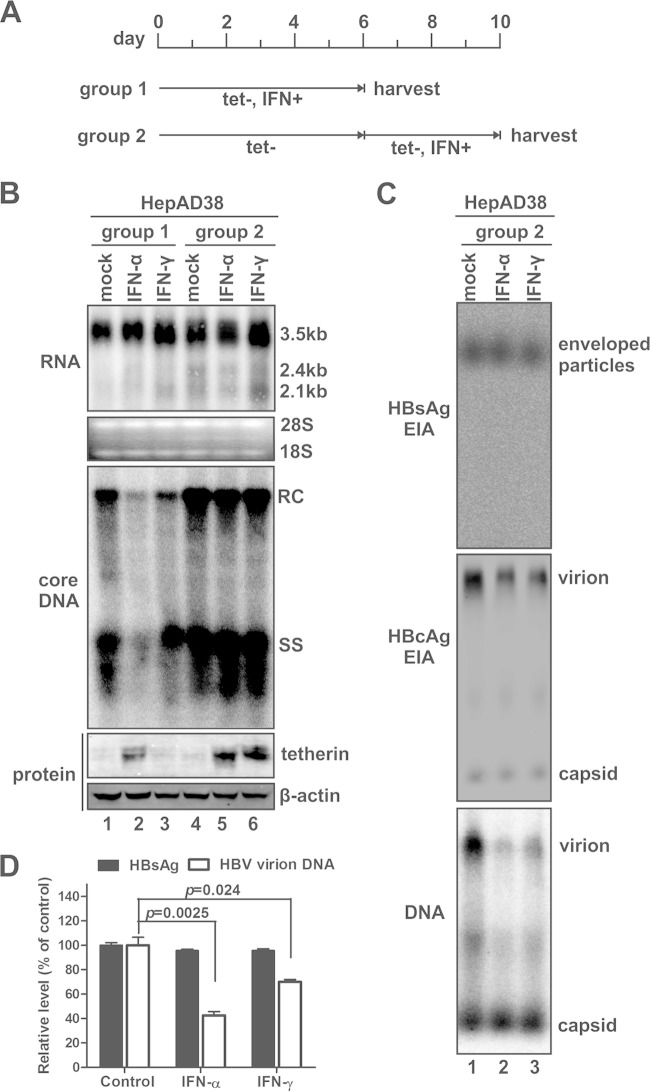
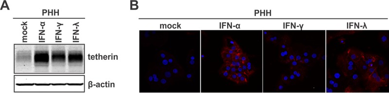
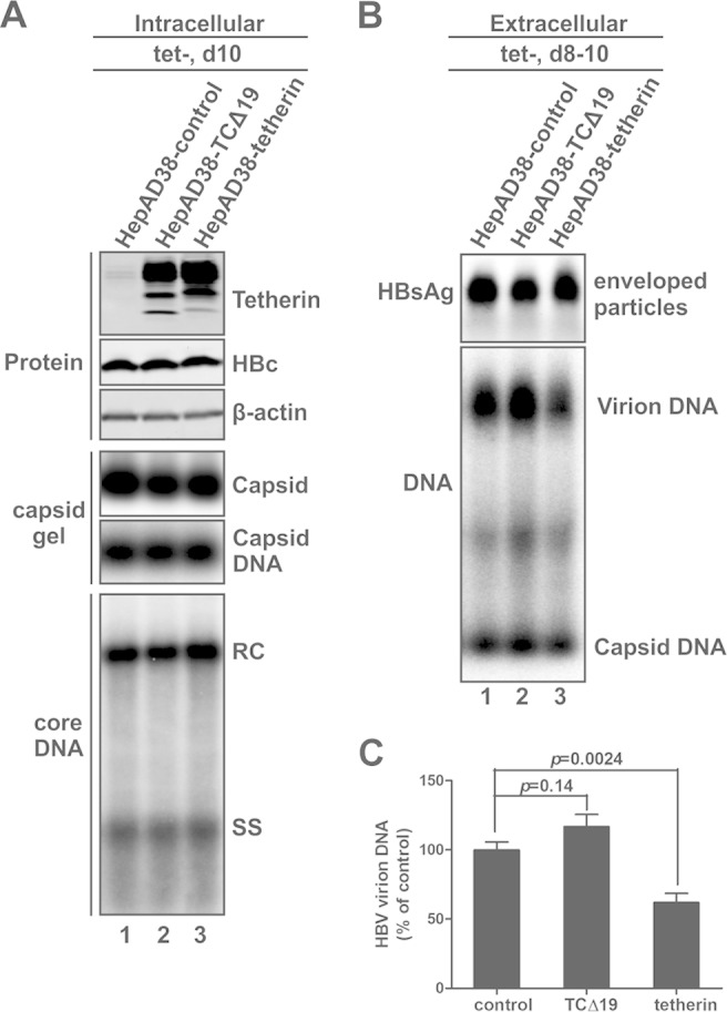
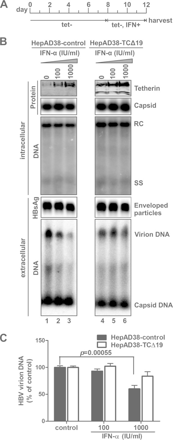
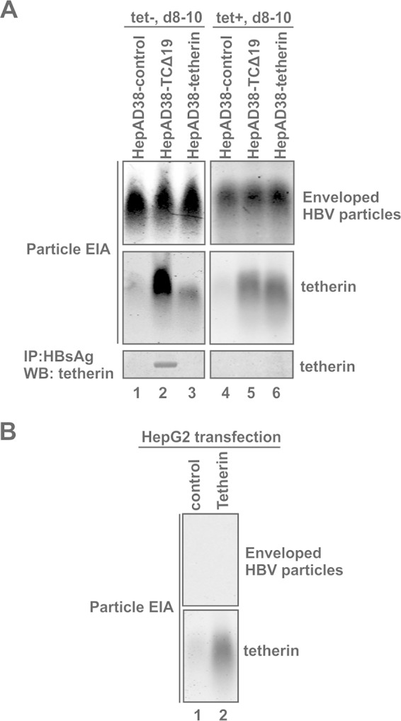
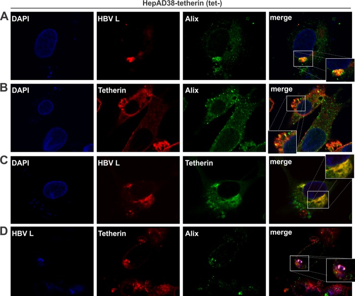
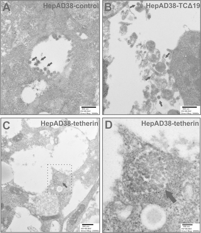
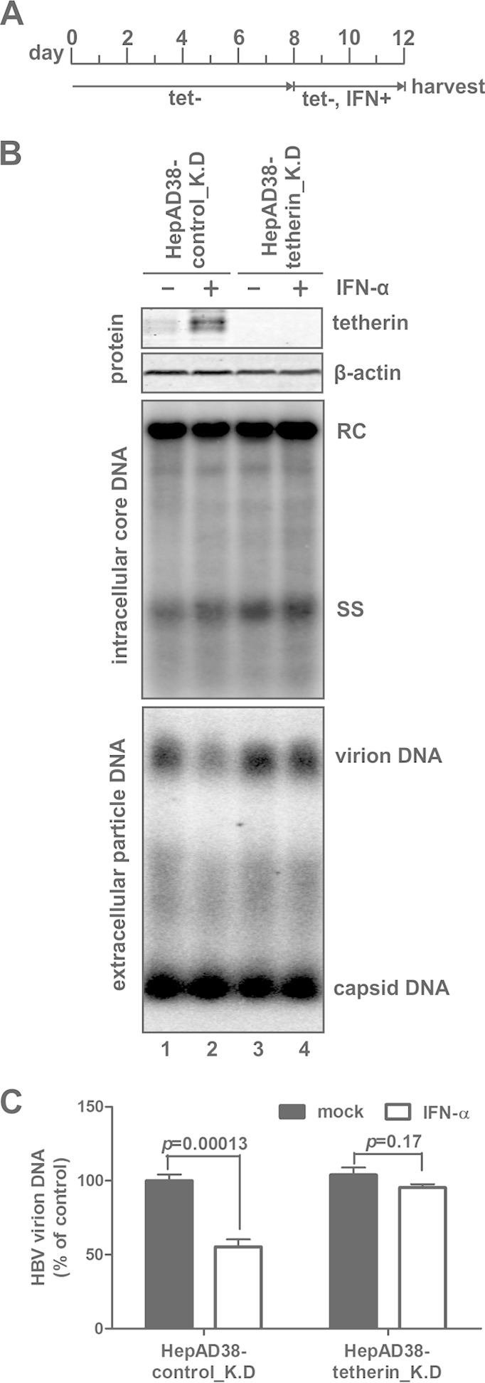
Interferon alpha (IFN-α) is an approved medication for chronic hepatitis B therapy. Besides acting as an immunomodulator, IFN-α elicits a pleiotropic antiviral state in hepatitis B virus (HBV)-infected hepatocytes, but whether or not IFN-α impedes the late steps of the HBV life cycle, such as HBV secretion, remains elusive. Here we report that IFN-α treatment of HepAD38 cells with established HBV replication selectively reduced HBV virion release without altering intracellular viral replication or the secretion of HBV subviral particles and nonenveloped capsids. In search of the interferon-stimulated gene(s) that is responsible for the reduction of HBV virion release, we found that tetherin, a broad-spectrum antiviral transmembrane protein that inhibits the egress of a variety of enveloped viruses, was highly induced by IFN-α in HepAD38 cells and in primary human hepatocytes. We further demonstrated that the expression of full-length tetherin, but not the C-terminal glycosylphosphatidylinositol (GPI) anchor-truncated form, inhibited HBV virion egress from HepAD38 cells. In addition, GPI anchor-truncated tetherin exhibited a dominant-negative effect and was incorporated into the liberated virions. We also found colocalization of tetherin and HBV L protein at the intracellular multivesicular body, where the budding of HBV virions takes place. In line with this, electron microscopy demonstrated that HBV virions were tethered in the lumen of the cisterna membrane under tetherin expression. Finally, knockdown of tetherin or overexpression of dominant negative tetherin attenuated the IFN-α-mediated reduction of HBV virion release. Taken together, our study suggests that IFN-α inhibits HBV virion egress from hepatocytes through the induction of tetherin.

Importance: Tetherin is a host restriction factor that blocks the egress of a variety of enveloped viruses through tethering the budding virions on the cell surface with its membrane anchor domains. Here we report that interferon directly and selectively inhibits the secretion of HBV virions, but not subviral particles or nonenveloped capsids, through the induction of tetherin in hepatocyte-derived cells. The antiviral function of tetherin requires the carboxyl-terminal GPI anchor, while the GPI anchor deletion mutant exhibits dominant negative activity and attaches to liberated HBV virions. Consistent with the fact that HBV is an intracellular budding virus, microscopy analyses demonstrated that the tethering of HBV virions occurs in the intracellular cisterna and that tetherin colocalizes with HBV virions on the multivesicular body, which is the HBV virion budding site. Our study not only expands the antiviral spectrum of tetherin but also sheds light on the mechanisms of interferon-elicited anti-HBV responses.


Figures

FIG 1
FIG 1
Interferon selectively inhibits HBV virion secretion in HepAD38 stable cells with established viral replication. (A) Schematic illustration of the experimental procedures. HepAD38 cells were cultured in a 6-well plate until confluent, and tetracycline (tet) was then removed from culture medium to induce HBV replication. One group of cells (group 1) was mock treated or treated with IFN-α (1,000 IU/ml) or IFN-γ (100 ng/ml) simultaneously with tetracycline withdrawal (tet−) for 6 days, and the cells and culture medium were then harvested; another group of cells (group 2) received interferon treatment after 6 days of HBV induction, and the treatment lasted 4 days before sample collection. Fresh culture media with or without IFN were replenished at 2-day intervals. (B) Cells harvested at the indicated time points were subjected to HBV RNA and core DNA analyses by Northern blotting and Southern blotting, respectively. rRNAs (28S and 18S) were used as total RNA loading controls. The positions of 3.5-kb and 2.4-kb/2.1-kb HBV RNAs and relaxed circular (RC) and single-stranded (SS) DNAs are indicated. The expression of tetherin was revealed by Western blotting, with β-actin serving as a loading control. (C) HBV particles in culture fluids harvested from group 2 cells at the endpoint of treatment (days 8 to 10) were analyzed by a particle gel assay. Enveloped HBV particles, including virions and subviral particles (HBsAg), were revealed by an EIA using HBsAb. Virion-associated capsids and nonenveloped capsids were detected by a core EIA. In situ-denatured HBV virion DNA and nonenveloped capsid DNA were detected by hybridization. (D) Culture fluid samples from group 2 were subjected to an HBsAg ELISA and HBsAg immunoprecipitation, and the immunoprecipitated samples were analyzed by HBV DNA qPCR. The relative levels of HBsAg and HBV DNA signals in each sample were plotted as a percentage of the signals from the mock-treated samples (means ± standard deviations).
FIG 2
FIG 2
Interferon induces tetherin expression in primary human hepatocytes (PHHs). PHHs were cultured in a 12-well plate and treated with IFN-α (1,000 IU/ml), IFN-γ (100 ng/ml), or IFN-λ (100 ng/ml) or left untreated (mock) for 2 days. The levels of tetherin expression were determined by a Western blot assay (A) and immunofluorescence microscopy (red) (B). β-Actin served as a Western blot loading control. Nuclei were stained with DAPI (blue).
FIG 3
FIG 3
Overexpression of wild-type but not GPI-anchor-deleted tetherin inhibits HBV virion release. Retroviral vector-transduced HepAD38 cells stably expressing wild-type or mutant tetherin, namely, HepAD38-tetherin and HepAD38-TCΔ19 cells, respectively, and control retrovirus-transduced HepAD38 cells (HepAD38-control) were cultured in complete maintenance medium until they became confluent, and tetracycline was then removed from the culture medium to induce HBV DNA replication. (A) Cells were harvested at day 10 after withdrawal of tetracycline, and intracellular tetherin expression, HBV core protein expression, capsid formation, and core DNA replication were analyzed. (B and C) The extracellular accumulation of HBV enveloped particles, virions, and naked capsid DNA from days 8 to 10 post-tetracycline induction was analyzed by a particle gel assay (B) and virion DNA IP-qPCR (C).
FIG 4
FIG 4
GPI-anchor-deleted tetherin (TCΔ19) rescues HBV virion egress with IFN-α treatment. (A) Experimental procedure. Confluent HepAD38-control and HepAD38-TCΔ19 cells were cultured in tetracycline-free medium for 8 days to stimulate HBV replication, and the cells were then mock treated or treated with IFN-α at the indicated concentrations for an additional 4 days, with medium changes and treatment being repeated every 2 days. (B) Harvested cell monolayers and culture fluids were subjected to intracellular and extracellular analyses, respectively, including Western blot analysis of tetherin expression and induction, a capsid gel EIA, core DNA Southern blot analysis, and a particle gel assay for enveloped HBV particles and particle-associated HBV DNA (from top to bottom). (C) The virion-associated DNA from 1 ml of the harvested supernatant was quantitated by HBsAg IP-qPCR and plotted as a percentage of the DNA signals from mock-treated cell samples (means ± standard deviations).
FIG 5
FIG 5
GPI-anchor-deleted tetherin is incorporated into secreted HBV virions. (A, top and middle) Culture medium of the indicated cell lines was collected between days 8 and 10 in the absence or presence of tetracycline and subjected to a particle gel assay, and the blot was stained for HBsAg and tetherin by specific antibodies. (Bottom) An aliquot of the culture fluid samples was subjected to HBsAg immunoprecipitation, followed by SDS-PAGE and Western blot analysis with tetherin antibodies. (B) HepG2 cells were transfected with the control vector or a plasmid expressing wild-type tetherin. Culture fluid was collected at day 5 posttransfection and subjected to a particle gel EIA for HBsAg and tetherin.
FIG 6
FIG 6
Intracellular colocalization of tetherin with HBV virions and MVBs. HepAD38-tetherin cells were cultured in tetracycline-free medium for 12 days and subjected to immunofluorescence confocal microscopy. (A) Colocalization of HBV virions (HBV L protein served as a marker) (red) and MVBs (Alix served as a marker) (green). Nuclei were counterstained with DAPI (blue). The inset in the merged image shows the colocalization signals (yellow) at a higher magnification. (B) Association between tetherin (red) and MVBs (Alix) (green). (C) Colocalization of HBV virions (L) (red) and tetherin (green). (D) Triple immunostaining of HBV virions (L) (blue), tetherin (red), and MVBs (Alix) (green). The colocalization signal (white) is further shown in the inset with an expanded view.
FIG 7
FIG 7
TEM of HBV virions. The indicated cells were cultured in tetracycline-free medium for 10 days, and ultrathin sections were prepared and negatively stained for TEM analysis. (A) HepAD38-control cells; (B) HepAD38-TCΔ19 cells; (C) HepAD38-tetherin cells; (D) expanded view of the area surrounded by a dotted line in panel C. Scale bars, high voltage (HV), and direct magnification are indicated. Representative HBV virions (∼40 to 50 nm) are indicated by arrows.
FIG 8
FIG 8
Knockdown of tetherin attenuates IFN-α-mediated suppression of HBV virion release. (A) Experimental procedure. HepAD38-control_K.D and HepAD38-tetherin_K.D cells were cultured in the presence of tetracycline until they became confluent, and the culture medium was then switched to tetracycline-free (tet−) medium to induce HBV replication. Eight days later, cells were untreated or treated with IFN-α (1,000 IU/ml) for another 4 days. During the entire culture period, fresh medium (with or without IFN-α) was replenished at 2-day intervals. (B) The harvested cell and supernatant samples were analyzed for tetherin expression, HBV core DNA replication, and extracellular accumulation of particle-associated HBV DNA. (C) The virion-associated DNA from 1 ml of the harvested supernatant was quantitated by HBsAg IP-qPCR and plotted as a percentage of the DNA signals from untreated HepAD38-control_K.D cell samples (means ± standard deviations).

Similar articles

Cited by

References

    1. Liang TJ. 2009. Hepatitis B: the virus and disease. Hepatology 49:S13–S21. doi:10.1002/hep.22881. - DOI - PMC - PubMed
    1. Block TM, Guo H, Guo JT. 2007. Molecular virology of hepatitis B virus for clinicians. Clin Liver Dis 11:685–706, vii. doi:10.1016/j.cld.2007.08.002. - DOI - PMC - PubMed
    1. Deny P, Zoulim F. 2010. Hepatitis B virus: from diagnosis to treatment. Pathol Biol (Paris) 58:245–253. doi:10.1016/j.patbio.2010.05.002. - DOI - PubMed
    1. Trepo C, Chan HL, Lok A. 2014. Hepatitis B virus infection. Lancet 384:2053–2063. doi:10.1016/S0140-6736(14)60220-8. - DOI - PubMed
    1. Seeger C, Mason WS. 2000. Hepatitis B virus biology. Microbiol Mol Biol Rev 64:51–68. doi:10.1128/MMBR.64.1.51-68.2000. - DOI - PMC - PubMed

Publication types

MeSH terms

LinkOut - more resources