

Primary combined hepatocellular-cholangiocellular sarcoma: An unusual case
- PMID: 26109824
- PMCID: PMC4476899
- DOI: 10.3748/wjg.v21.i23.7335
Primary combined hepatocellular-cholangiocellular sarcoma: An unusual case
Abstract
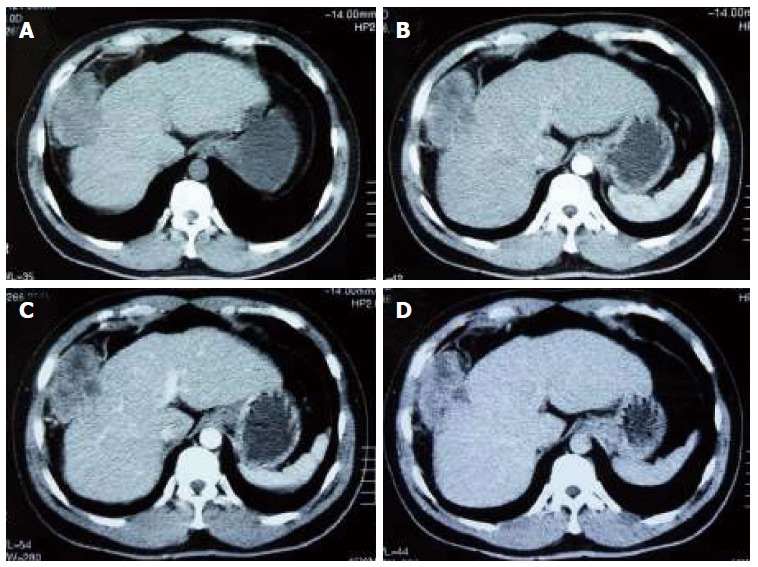
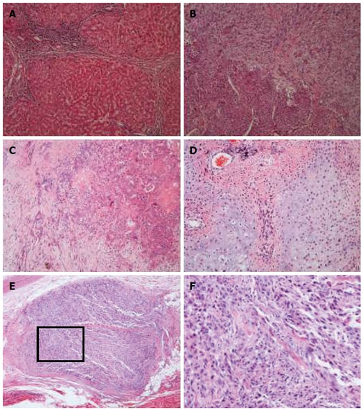
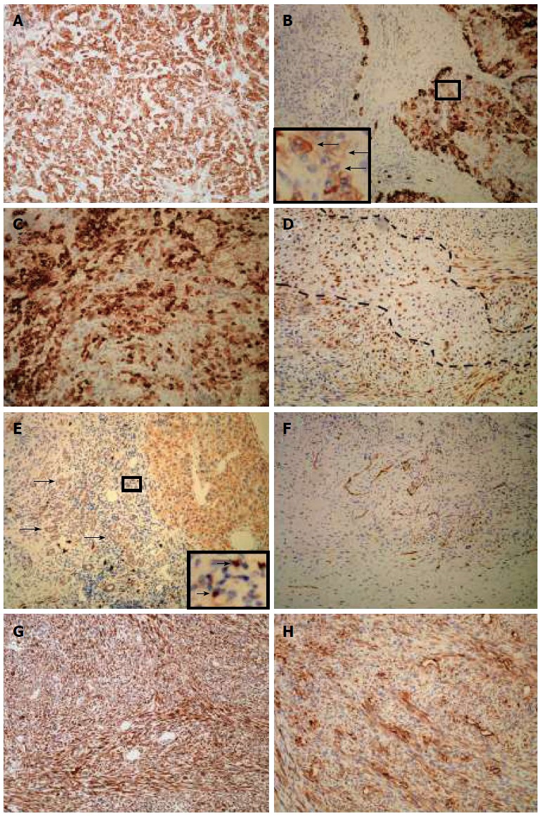
Primary liver carcinosarcoma is rare. Here we report an unusual case of liver carcinosarcoma containing combined hepatocellular cholangiocarcinoma. A mass in the right liver lobe of a 45-year-old man was accidentally discovered by ultrasonic inspection and computed tomography (CT) scan. Surgical resection was performed following a diagnosis of primary liver cancer. Micropathologically, both carcinomatous and sarcomatous elements were present, and diagnosis of liver carcinosarcoma was confirmed. The carcinomatous element consisted of hepatocellular carcinoma and foci of cholangiocellular carcinoma. The sarcomatous element was composed of spindle cells and bizarre cells, as well as foci of osteosarcoma and chondrosarcoma. Hepatocellular carcinoma cells diffusely expressed both hepatocyte specific markers cytokeratin (CK) 8/18 and cholangiocyte specific markers CK19, and sarcoma cells were positive for vimentin. Interestingly, both carcinomatous and sarcomatous cells expressed epithelial membrane antigen. CD117-positive ductular reactions and small undifferentiated cells were observed. A liver progenitor cell origin of the liver carcinosarcoma was proposed.
Keywords: Carcinosarcoma; Cholangiocellular carcinoma; Hepatocellular carcinoma; Liver neoplasm; Stem cells.
Figures
References
-
- Nakajima T, Kubosawa H, Kondo Y, Konno A, Iwama S. Combined hepatocellular-cholangiocarcinoma with variable sarcomatous transformation. Am J Clin Pathol. 1988;90:309–312. - PubMed
-
- Goto H, Tanaka A, Kondo F, Takeshita K, Nagashima I, Hanawa N, Aiso M, Takamori Y, Kato K, Takahashi Y, et al. Carcinosarcoma of the liver. Intern Med. 2010;49:2577–2582. - PubMed
-
- Papotti M, Sambataro D, Marchesa P, Negro F. A combined hepatocellular/cholangiocellular carcinoma with sarcomatoid features. Liver. 1997;17:47–52. - PubMed
-
- Durnez A, Verslype C, Nevens F, Fevery J, Aerts R, Pirenne J, Lesaffre E, Libbrecht L, Desmet V, Roskams T. The clinicopathological and prognostic relevance of cytokeratin 7 and 19 expression in hepatocellular carcinoma. A possible progenitor cell origin. Histopathology. 2006;49:138–151. - PubMed
Publication types
MeSH terms
Substances
LinkOut - more resources
Full Text Sources
Other Literature Sources
Medical
Research Materials
