

Neuroprotective effects of ginsenoside Rg1-induced neural stem cell transplantation on hypoxic-ischemic encephalopathy
- PMID: 26109949
- PMCID: PMC4468766
- DOI: 10.4103/1673-5374.156971
Neuroprotective effects of ginsenoside Rg1-induced neural stem cell transplantation on hypoxic-ischemic encephalopathy
Abstract
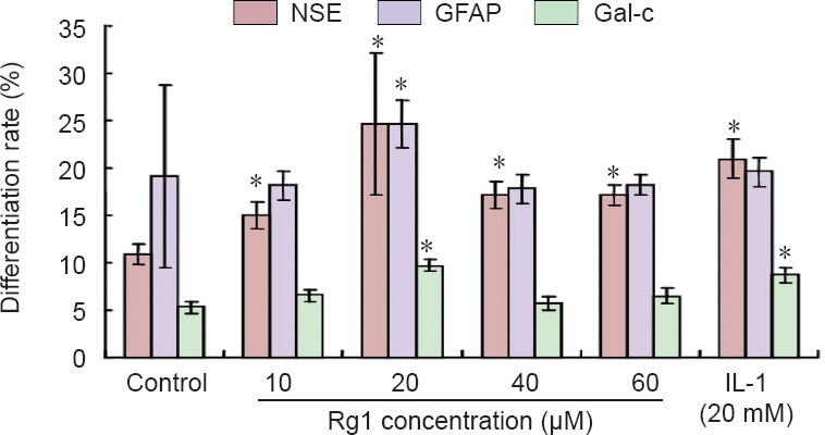
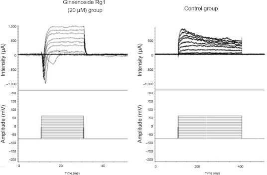
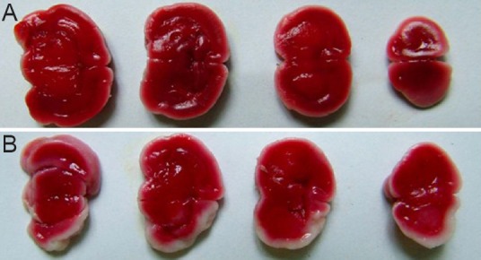
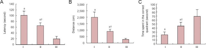
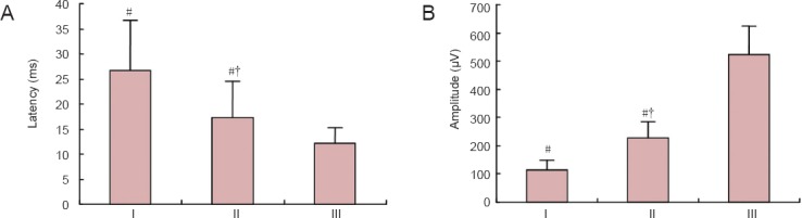
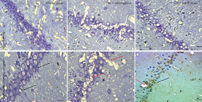
Ginsenoside Rg1 is the major pharmacologically active component of ginseng, and is reported to have various therapeutic actions. To determine whether it induces the differentiation of neural stem cells, and whether neural stem cell transplantation after induction has therapeutic effects on hypoxic-ischemic encephalopathy, we cultured neural stem cells in 10-80 μM ginsenoside Rg1. Immunohistochemistry revealed that of the concentrations tested, 20 mM ginsenoside Rg1 had the greatest differentiation-inducing effect and was the concentration used for subsequent experiments. Whole-cell patch clamp showed that neural stem cells induced by 20 μM ginsenoside Rg1 were more mature than non-induced cells. We then established neonatal rat models of hypoxic-ischemic encephalopathy using the suture method, and ginsenoside Rg1-induced neural stem cells were transplanted via intracerebroventricular injection. These tests confirmed that neural stem cells induced by ginsenoside had fewer pathological lesions and had a significantly better behavioral capacity than model rats that received saline. Transplanted neural stem cells expressed neuron-specific enolase, and were mainly distributed in the hippocampus and cerebral cortex. The present data suggest that ginsenoside Rg1-induced neural stem cells can promote the partial recovery of complicated brain functions in models of hypoxic-ischemic encephalopathy.
Keywords: cell differentiation; cell transplantation; cognition; ginsenoside Rg1; hypoxic-ischemic brain damage; nerve reconstruction; nerve regeneration; neural regeneration; neural stem cells.
Conflict of interest statement
Figures
References
-
- Abend NS, Licht DJ. Predicting outcome in children with hypoxic ischemic encephalopathy. Pediatr Crit Care Med. 2008;9:32–39. - PubMed
-
- Andsberg G, Björklund ZKa, Lindvall O, Martínez-Serrano A. Amelioration of ischaemia-induced neuronal death in the rat striatum by NGF-secreting neural stem cells. Eur J Neurosci. 1998;10:2026–2036. - PubMed
-
- Carrai R, Grippo A, Lori S, Pinto F, Amantini A. Prognostic value of somatosensory evoked potentials in comatose children: a systematic literature review. Intensive Care Med. 2010;36:1112–1126. - PubMed
-
- Dive D, Giffroy X. Somatosensory evoked potentials: clinical applications in peripheral neuropathies. Rev Med Liege. 2004;59(Suppl 1):157–169. - PubMed
LinkOut - more resources
Full Text Sources
Other Literature Sources