. 2015 Jun 25;10(6):e0130653.
doi: 10.1371/journal.pone.0130653. eCollection 2015.

Visual vs Fully Automatic Histogram-Based Assessment of Idiopathic Pulmonary Fibrosis (IPF) Progression Using Sequential Multidetector Computed Tomography (MDCT)

Affiliations

Visual vs Fully Automatic Histogram-Based Assessment of Idiopathic Pulmonary Fibrosis (IPF) Progression Using Sequential Multidetector Computed Tomography (MDCT)

Davide Colombi et al. PLoS One. .

Abstract

Objectives: To describe changes over time in extent of idiopathic pulmonary fibrosis (IPF) at multidetector computed tomography (MDCT) assessed by semi-quantitative visual scores (VSs) and fully automatic histogram-based quantitative evaluation and to test the relationship between these two methods of quantification.

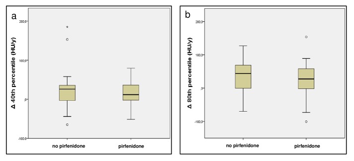
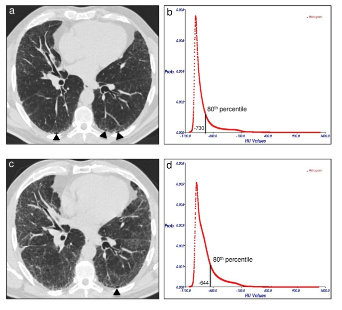
Methods: Forty IPF patients (median age: 70 y, interquartile: 62-75 years; M:F, 33:7) that underwent 2 MDCT at different time points with a median interval of 13 months (interquartile: 10-17 months) were retrospectively evaluated. In-house software YACTA quantified automatically lung density histogram (10th-90th percentile in 5th percentile steps). Longitudinal changes in VSs and in the percentiles of attenuation histogram were obtained in 20 untreated patients and 20 patients treated with pirfenidone. Pearson correlation analysis was used to test the relationship between VSs and selected percentiles.

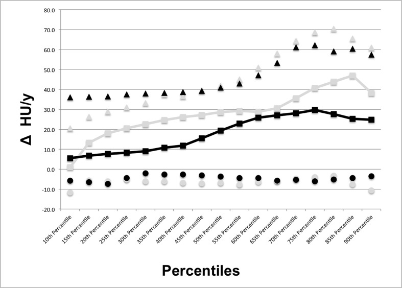
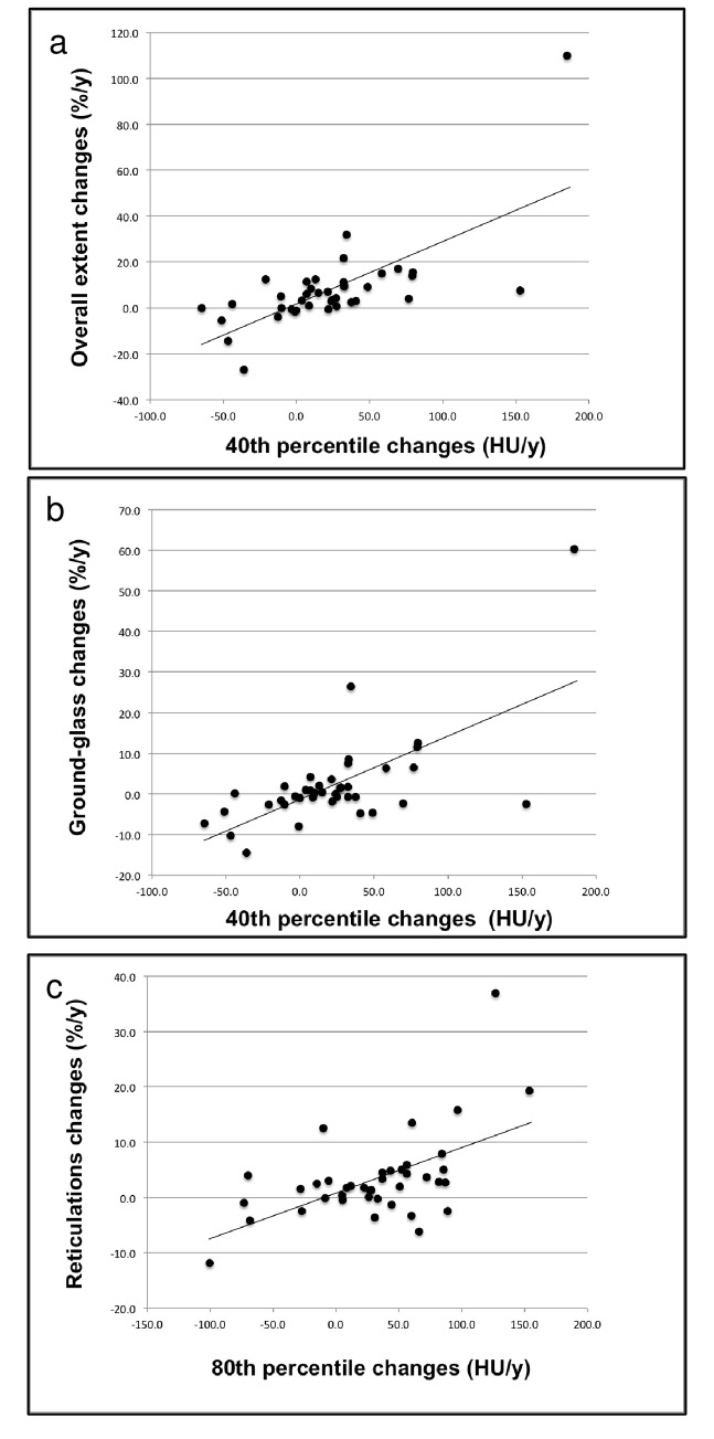
Results: In follow-up MDCT, visual overall extent of parenchymal abnormalities (OE) increased in median by 5%/year (interquartile: 0%/y; +11%/y). Substantial difference was found between treated and untreated patients in HU changes of the 40th and of the 80th percentiles of density histogram. Correlation analysis between VSs and selected percentiles showed higher correlation between the changes (Δ) in OE and Δ 40th percentile (r=0.69; p<0.001) as compared to Δ 80th percentile (r=0.58; p<0.001); closer correlation was found between Δ ground-glass extent and Δ 40th percentile (r=0.66, p<0.001) as compared to Δ 80th percentile (r=0.47, p=0.002), while the Δ reticulations correlated better with the Δ 80th percentile (r=0.56, p<0.001) in comparison to Δ 40th percentile (r=0.43, p=0.003).

Conclusions: There is a relevant and fully automatically measurable difference at MDCT in VSs and in histogram analysis at one year follow-up of IPF patients, whether treated or untreated: Δ 40th percentile might reflect the change in overall extent of lung abnormalities, notably of ground-glass pattern; furthermore Δ 80th percentile might reveal the course of reticular opacities.


Conflict of interest statement

Competing Interests: Colombi D received a support for travel to meetings for the study from Intermune. Oltmanns U received payment for educational presentation from European Respiratory Society and fees for both meetings and presentation at congresses from Intermune. Palmowski K received meetings expenses from Intermune. Herth F is board member of Novartis, Astra Zeneca, Grifols, Berlin Chemie, Chiesi, Pulmonx, and PneumRx; he also received fees for lectures from the same companies. Kauczor HU is board member of Siemens; he also received fees for lectures and educational presentations from Bracco, Novartis, Siemens, Boehringer, and Bayer. Sverzellati N received fees for lectures and educational presentation from Intermune, Boeringher, Astrazeneca, Pfizer, and Chiesi. Kreuter M received a grant from Dietmar Hopp Stiftung; he is also a consultant for Intermune and he received fees for lectures and educational presentations from Boehringer and Intermune. Heussel CP is a consultant of Pfizer, Boheringer Ingelheim, Gilead, Intermune, and Fresenius; he also received research funding from Siemens, Pfizer, and Boheringer Ingelheim as well as fees for lectures from Gilead, MSD, Pfizer, Intermune, Boheringer Ingelheim, and Novartis. This does not alter the authors' adherence to PLOS ONE policies on sharing data and materials. For the remaining authors no competing interests were declared.

Figures

Fig 1
Fig 1. Visualization of the fully automatic lung parenchyma segmentation as obtained by in-house YACTA software.
Sagittal reconstruction image of a non-enhanced MDCT scan obtained from a patient suffering from idiopathic pulmonary fibrosis (IPF) not included in the current trial. YACTA software automatically segmented lung parenchyma and trachea-bronchial tree, emphasized as green and orange overlay respectively (window width: 1600 HU; level: -600 HU). Note that the segmentation algorithm fails to segment portions of the lung parenchyma in the sub-pleural space of the recessus, due to its similar density to the chest wall. (MDCT = multidetector computed tomography).
Fig 2
Fig 2. Median density changes at 1-year follow-up in the range of 10th-90th percentile of the MDCT attenuation histogram for patients suffering from idiopathic pulmonary fibrosis (IPF) treated and untreated with pirfenidone.
The largest difference with the lowest overlap between treated and untreated patients in longitudinal HU changes of the attenuation histogram was detected in the 40th and in the 80th percentiles. Black squares = median increase of each 5th percentile step included in the 10th-90th range of the lung density histogram for patients treated with pirfenidone; black triangles and circles = interquartile range of the increase in each 5th percentile step included in the 10th-90th range of the lung density histogram for patients treated with pirfenidone; grey squares = median increase of each 5th percentile step included in the 10th-90th range of the lung density histogram for patients not treated with pirfenidone; grey triangles and circles = interquartile range of the increase in each 5th percentile step included in the 10th-90th range of the lung density histogram for patients not treated with pirfenidone. (Δ HU/y = changes in Hounsfield units per year; MDCT = multidetector computed tomography).
Fig 3
Fig 3. Distribution of the density changes at 1-year in the selected percentiles from the MDCT attenuation histogram of patients suffering from idiopathic pulmonary fibrosis (IPF) treated and untreated with pirfenidone.
Box and Whisker plots represent the 40th percentile (a) and the 80th percentile distributions (b) of lung density histogram as observed in patients treated with pirfenidone and in patients not treated with pirfenidone. The central line represents the median, the yellow box encompasses the 25th-75th percentiles, whiskers show the 10th-90th percentile, and the empty circles represent individual outliers. (HU/y = Hounsfield units per year; Δ 40th percentile = changes at 1-year in the 40th percentile of the attenuation histogram; Δ 80th percentile = changes at 1-year in the 80th percentile of the attenuation histogram; MDCT = multidetector computed tomography).
Fig 4
Fig 4. Correlations between changes at 1-year in selected percentiles of MDCT attenuation histogram and visual scores.
Dot plots with linear regression curves for changes in overall extent of parenchymal abnormalities plotted against density variation in the 40th percentile of the MDCT attenuation histogram (r = 0.69, p < 0.001) (a), for the changes in ground-glass opacity extent plotted against density variation in the 40th percentile of the MDCT attenuation histogram (r = 0.66, p < 0.001) (b), and for changes in reticulations extent plotted against density variation in 80th percentile of the MDCT attenuation histogram (r = 0.56, p < 0.001) (c). (%/y = percent per year; HU/y = Hounsfield units per year; MDCT = multidetector computed tomography).
Fig 5
Fig 5. Variation of disease extent at follow-up MDCT in a 53 years-old man suffering from idiopathic pulmonary fibrosis (IPF) not treated with pirfenidone.
Axial MDCT image at level 5 (1 cm above the right hemi-diaphragm as described in the text) of the initial examination shows mild reticular opacities and minimal honeycombing (arrowheads) in a sub-pleural distribution (a). The 80th percentile of the initial MDCT attenuation histogram corresponded to -730 HU (b). Follow-up MDCT scan at the same axial level after 17 months shows a predominant increase of reticulations (visual score = +22%) and unmodified honeycombing extent (arrowhead) (c). An increase of 86 HU in the density of the 80th percentile was seen between initial and follow-up MDCT attenuation histograms (d). (Prob. = probability; HU = Hounsfield units; MDCT = multidetector computed tomography).

Similar articles

Cited by

References

    1. Raghu G, Collard HR, Egan JJ, Martinez FJ, Behr J, Brown KK, et al. An official ATS/ERS/JRS/ALAT statement: idiopathic pulmonary fibrosis: evidence-based guidelines for diagnosis and management. Am J Respir Crit Care Med 2011;183: 788–824. 10.1164/rccm.2009-040GL - DOI - PMC - PubMed
    1. King TE Jr, Pardo A, Selman M. Idiopathic pulmonary fibrosis. Lancet 2011;378: 1949–1961. 10.1016/S0140-6736(11)60052-4 - DOI - PubMed
    1. Kreuter M. Pirfenidone: an update on clinical trial data and insights from everyday practice. Eur Respir Rev 2014;23: 111–117. 10.1183/09059180.00008513 - DOI - PMC - PubMed
    1. King TE Jr, Bradford WZ, Castro-Bernardini S, Fagan EA, Glaspole I, Glassberg MK, et al. A phase 3 trial of pirfenidone in patients with idiopathic pulmonary fibrosis. N Engl J Med 2014;370: 2083–2092. 10.1056/NEJMoa1402582 - DOI - PubMed
    1. Richeldi L, du Bois RM, Raghu G, Azuma A, Brown KK, Costabel U, et al. Efficacy and safety of nintedanib in idiopathic pulmonary fibrosis. N Engl J Med 2014;370: 2071–2082. 10.1056/NEJMoa1402584 - DOI - PubMed

Publication types

LinkOut - more resources