. 2015 Jun 25;11(6):e1005167.
doi: 10.1371/journal.pgen.1005167. eCollection 2015 Jun.

AKTIP/Ft1, a New Shelterin-Interacting Factor Required for Telomere Maintenance

Affiliations

AKTIP/Ft1, a New Shelterin-Interacting Factor Required for Telomere Maintenance

Romina Burla et al. PLoS Genet. .

Abstract

Telomeres are nucleoprotein complexes that protect the ends of linear chromosomes from incomplete replication, degradation and detection as DNA breaks. Mammalian telomeres are protected by shelterin, a multiprotein complex that binds the TTAGGG telomeric repeats and recruits a series of additional factors that are essential for telomere function. Although many shelterin-associated proteins have been so far identified, the inventory of shelterin-interacting factors required for telomere maintenance is still largely incomplete. Here, we characterize AKTIP/Ft1 (human AKTIP and mouse Ft1 are orthologous), a novel mammalian shelterin-bound factor identified on the basis of its homology with the Drosophila telomere protein Pendolino. AKTIP/Ft1 shares homology with the E2 variant ubiquitin-conjugating (UEV) enzymes and has been previously implicated in the control of apoptosis and in vesicle trafficking. RNAi-mediated depletion of AKTIP results in formation of telomere dysfunction foci (TIFs). Consistent with these results, AKTIP interacts with telomeric DNA and binds the shelterin components TRF1 and TRF2 both in vivo and in vitro. Analysis of AKTIP- depleted human primary fibroblasts showed that they are defective in PCNA recruiting and arrest in the S phase due to the activation of the intra S checkpoint. Accordingly, AKTIP physically interacts with PCNA and the RPA70 DNA replication factor. Ft1-depleted p53-/- MEFs did not arrest in the S phase but displayed significant increases in multiple telomeric signals (MTS) and sister telomere associations (STAs), two hallmarks of defective telomere replication. In addition, we found an epistatic relation for MST formation between Ft1 and TRF1, which has been previously shown to be required for replication fork progression through telomeric DNA. Ch-IP experiments further suggested that in AKTIP-depleted cells undergoing the S phase, TRF1 is less tightly bound to telomeric DNA than in controls. Thus, our results collectively suggest that AKTIP/Ft1 works in concert with TRF1 to facilitate telomeric DNA replication.


Conflict of interest statement

The authors have declared that no competing interests exist.

Figures
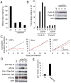
Fig 1
Fig 1. AKTIP depletion affects cell cycle progression and induces the DNA damage response (DDR).
(A) Relative mitotic indexes (± SD) of shAKTIP (hairpin sequences 9, 11 and 13) and control (ctr) at 10 dpi. (B) Immunoblotting of 10 dpi extracts from shAKTIP HPFs shows accumulation of cyclins compared to ctr; in the graph, blot signals are normalized relative to the actin used as a loading control. (C) Cumulative population doublings of HPFs, HeLa and 293T cells transduced with ctr or shAKTIP-11. (D) Immunoblots of 10 dpi extracts reveal DNA damage signaling in shAKTIP samples. Densitometric analysis showed that in 9, 11 and 13 RNAi cells there is a 2.7-, 2.6-, and 3.3-fold increase of Chk1, respectively; in the same cells ATM-P Ser 1981 increases were 1.4-, 1.6- and 2.0-fold, respectively. (E) Q-PCR of total RNA shows a strong increase in p21 expression in shAKTIP-11 HPFs relative to control; samples collected at 7 dpi were analyzed in duplicate and shown as mean values ± SD. See also S1 Fig.
Fig 2
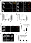
Fig 2. AKTIP downregulation induces TIF formation.
(A) γH2AX (green in merges), ATM-P (red in merges) and 53BP1 (red in merges) foci in 5 dpi shAKTIP-11 HPFs. Foci are absent in mock and 5 dpi ctr HPFs. (B, C) Numbers of foci per cell (B) and percents of cells with more than 5 foci (C); bars are the mean values from two independent experiments ± SD. (D) TIFs in 5 dpi shAKTIP-11 HPFs (in merges, γH2AX is green and TRF1 red); arrowheads point to TIFs. X-ray-treated (IR; 1 Gy) HPFs were used as controls. (E) Percents of γH2AX foci co-localizing with TRF1 (TIFs), and of cells with more than 5 TIFs. Bars are the mean values from two independent experiments ± SD. ***, **, * indicate p<0.001 p<0.01 p<0.05 in the Student t test, respectively. See also S2 and S3 Figs. (F-G) Depletion of the AKT kinase does not induce formation of 53BP1 DNA repair foci. (F) Examples of 53BP1 foci in mock, ctr, and shAKT HPFs. (G) Quantification of 53BP1 foci in mock, ctr, and shAKT HPFs. 100 cells scored for each sample. Values are the means of two independent experiments ± SD, and are not significantly different in the Student t test.
Fig 3
Fig 3. Ft1 downregulation leads to telomeric aberrations.
(A) Partial DAPI-stained (red) metaphases from ctr or shFt1 MEFs showing telomeric FISH signals (black and white; green in merges); arrows indicate selected multiple telomeric signals (MTSs), and arrowhead points to sister telomere associations (STAs). (B) MTS, STA and telomere fusion (TF) frequencies in mock and 7 dpi ctr and shFt1 MEFs; *** significantly different from controls in the χ2 test with p<0.001. Values are the mean frequencies from 2 independent experiments. (C) Trf1 is epistatic to Ft1. The MTS frequencies observed in shFt1, shTrf1 and shFt1 + shTrf1 are not significantly different in the χ2 test, but the STA frequencies observed in shFt1, shTrf1 and shFt1 + shTrf1 are significantly higher that that seen shFt1 (***different in the χ2 test with p<0.001). Values are the mean frequencies from 2 independent experiments. (D) Relative frequencies of the indicated chromosome types; frequencies were calculated from pooled data (B and C). (E) Observed (O) and expected (E; on the basis of independence; calculated from pooled data using the binomial distribution formula) frequencies of sister telomere pairs with the indicated FISH patterns; *** O-E differences significant with p<0.001 in the χ2 test. See also S4 and S5 Figs.
Fig 4
Fig 4. AKTIP interacts with telomeric DNA, TRF1 and TRF2.
(A) ChIPs from HPFs, uninfected HeLa cells and shAKTIP-11-infected HeLa cells reveal interactions between AKTIP and telomeric DNA. Chromatin was immunoprecipitated with an anti-AKTIP antibody or control IgGs; slot-blots were hybridized with TTAGGG or ALU repeat probes. (B) ChIP quantification after normalization to the input (levels shown in A). Bars show the mean values of two experiments ± SD; the amount of telomeric DNA precipitated from uninfected HeLa cells is significantly higher than that obtained from shAKTIP-11-infected cells (*p<0.05 in the Student t test). (C, D) AKTIP-GST pulls down TRF1 (C) and TRF2 (D) from 293T cell extracts. In, input; M, MW markers.
Fig 5
Fig 5. AKTIP directly binds TRF1 and TRF2.
(A) A tridimensional molecular model for AKTIP. The arrows point to the starting sites of the disordered N- and C-terminal regions (not depicted); the variant Asp residue and His-Pro-Leu motif are represented as sticks and indicated by red and purple arrows, respectively (see also S5 Fig). (B) Schematic organization of the AKTIP protein; the AKTIP truncations used for GST pulldown are indicated below the scheme. (C, D) In vitro mapping the AKTIP regions that interact with TRF1 or TRF2 using bacterially purified proteins; the UEV domain of AKTIP binds both TRF1 and TRF2.
Fig 6
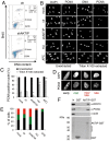
Fig 6. AKTIP downregulation impairs DNA replication.
(A) FACS analysis of 10 dpi ctr and shAKTIP-11 HPFs incubated with BrdU for 30 min, fixed, and then stained for BrdU and DNA (with PI). AKTIP depletion results in an S phase block; percents of cells in different cell cycle phases are reported in the upper right corner of each panel. (B, C) PCNA localization in unsynchronized mock, 10 dpi ctr, 10 dpi shAKTIP-11, HU-treated and APC-treated HPFs. Examples (B) and quantification of PCNA positive nuclei (C) from unextracted or Triton X-100-extracted HPFs. Bars are the mean values ± SD of samples analyzed in duplicate. ** and * indicate significant difference from control with p<0.01 and p<0.05 in the Student t test. (D, E) Distribution of nuclei with different S phase PCNA staining patterns. Bars in the graph (E) are the mean values ± SD of samples analyzed in duplicate; colours in E are as in the representative images shown in D; distributions of ctr and shAKTIP nuclei are significantly different in the Student t test with p<0.05. (F) AKTIP-GST pulls down PCNA and RPA70 from 293T cell extracts.
Fig 7
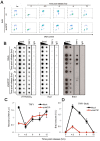
Fig 7. AKTIP downregulation impairs telomere replication.
Ctr or shAKTIP HeLa cells were synchronized at the G1/S boundary with a double thymidine block and harvested at the indicated times. (A) Scatter plots showing the proportions of cells in S phase in asynchronous cultures (As) and in cultures analyzed at various times after release from the double thymidine block. Prior to harvest at each time point, the cells were incubated with BrdU for 30 min. (B) ChIP analysis on synchronized HeLa cells incubated with BrdU for 1 h before harvesting. Precipitations were performed with an anti-TRF1 antibody. IgG antibody was used as negative control. Inputs represent 10 and 1% of genomic DNA. Dot-blot analysis was performed using telomeric or ALU repeat-specific probes. Precipitated DNA was analyzed by Western blotting with an anti-BrdU antibody. (C, D) Quantification of the data expressed in arbitrary units (A.U.) of unlabeled (C) or BrdU-labeled (D) precipitated telomeric DNA at the different time points of analysis, each normalized to input samples. The graphs show three independent experiments, with error bars indicating the SD.
Fig 8
Fig 8. AKTIP localizes at the nuclear periphery.
(A) Immunolocalization of AKTIP in HPFs and HeLa cells with an anti-AKTIP antibody, and in AKTIP-FLAG expressing 293T cells with an anti-FLAG antibody. shAKTIP HPFs show a strong reduction of the AKTIP signal. (B) Optical sections of a HPF and a HeLa cell showing AKTIP enrichment at the nuclear periphery. (C) Co-immunostaining of detergent-extracted HPFs for AKTIP and TRF1 (projection of 8 z stacks) showing a limited signal co-localization.

Similar articles

Cited by

References

    1. Blackburn E, Greider C, Szostak J. Telomeres and telomerase: the path from maize, Tetrahymena and yeast to human cancer and aging. Nat Med. 2006;12(10):1133–8. - PubMed
    1. Palm W, de Lange T. How shelterin protects mammalian telomeres. Ann Rev Genet. 2008;42:301–34. 10.1146/annurev.genet.41.110306.130350 - DOI - PubMed
    1. Hockemeyer D, Sfeir A, Shay J, Wright W, de Lange T. POT1 protects telomeres from a transient DNA damage response and determines how human chromosomes end. Embo J. 2005;24(14):2667–78. - PMC - PubMed
    1. Hockemeyer D, Palm W, Else T, Daniels J, Takai K, Ye J, et al. Telomere protection by mammalian Pot1 requires interaction with Tpp1. Nat Struct Mol Biol. 2007;14(8):754–61. - PubMed
    1. Denchi E, de Lange T. Protection of telomeres through independent control of ATM and ATR by TRF2 and POT1. Nature. 2007;448:1068–71. - PubMed

Publication types

MeSH terms