

Extracts of Feijoa Inhibit Toll-Like Receptor 2 Signaling and Activate Autophagy Implicating a Role in Dietary Control of IBD
- PMID: 26110654
- PMCID: PMC4482396
- DOI: 10.1371/journal.pone.0130910
Extracts of Feijoa Inhibit Toll-Like Receptor 2 Signaling and Activate Autophagy Implicating a Role in Dietary Control of IBD
Abstract
Background: Inflammatory bowel disease (IBD) is a heterogeneous chronic inflammatory disease affecting the gut with limited treatment success for its sufferers. This suggests the need for better understanding of the different subtypes of the disease as well as nutritional interventions to compliment current treatments. In this study we assess the ability of a hydrophilic feijoa fraction (F3) to modulate autophagy a process known to regulate inflammation, via TLR2 using IBD cell lines.
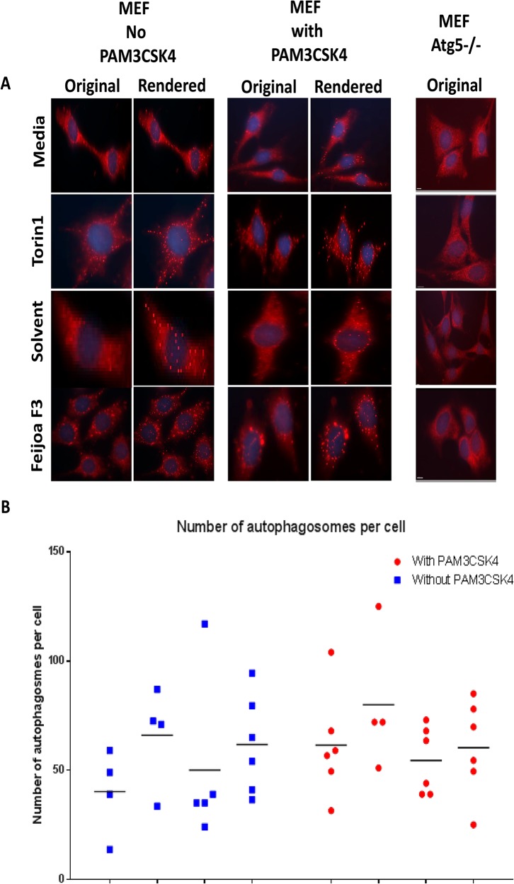
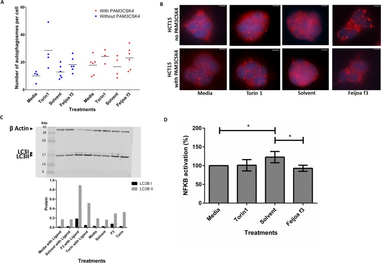
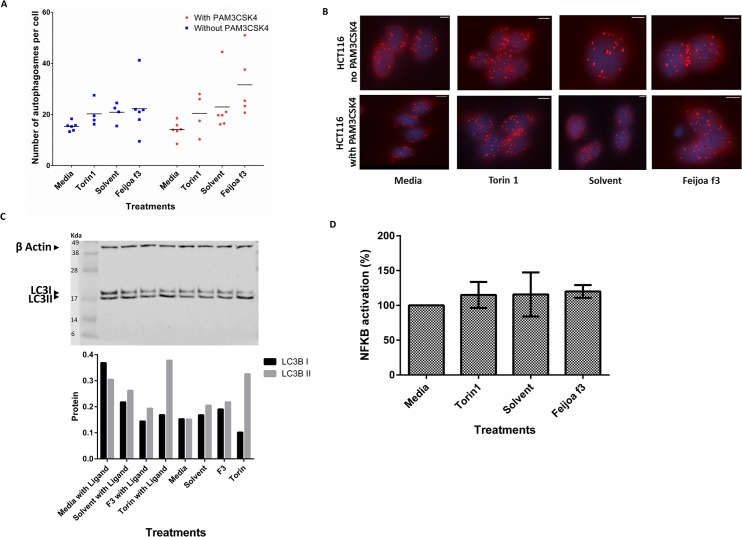
Method: Mouse embryonic fibroblasts (MEF) deleted for ATG5, and two intestinal epithelial cells HCT15 and HCT116, were used to test the anti-inflammatory effect of F3 after stimulating the cells with a TLR2 specific ligand PAM3CSK4.
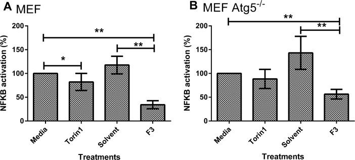
Results: F3 was able to reduce TLR2 specific inflammation and stimulate autophagy in MEFs and HCT15 cells but not in HCT116 cells. The anti-inflammatory effect was reduced in the MEF cells deleted for ATG5. In addition, the activation of autophagy by F3 was enhanced by PAM3CSK4.
Conclusion: F3 of feijoa can interact with cells via a TLR2 specific mechanism and reduce Nuclear factor kappa B (NF-κB) activation in part due to stimulation of autophagy. These results suggest that there is potential benefit in using feijoa extracts as part of dietary interventions to manage IBD in patients.
Conflict of interest statement
Figures
Similar articles
-
Screening of Cytotoxicity and Anti-Inflammatory Properties of Feijoa Extracts Using Genetically Modified Cell Models Targeting TLR2, TLR4 and NOD2 Pathways, and the Implication for Inflammatory Bowel Disease.Nutrients. 2018 Aug 31;10(9):1188. doi: 10.3390/nu10091188. Nutrients. 2018. PMID: 30200338 Free PMC article.
-
Anti-inflammatory activity of fruit fractions in vitro, mediated through toll-like receptor 4 and 2 in the context of inflammatory bowel disease.Nutrients. 2014 Nov 19;6(11):5265-79. doi: 10.3390/nu6115265. Nutrients. 2014. PMID: 25415606 Free PMC article.
-
HIC1 Tumor Suppressor Loss Potentiates TLR2/NF-κB Signaling and Promotes Tissue Damage-Associated Tumorigenesis.Mol Cancer Res. 2015 Jul;13(7):1139-48. doi: 10.1158/1541-7786.MCR-15-0033. Epub 2015 May 1. Mol Cancer Res. 2015. PMID: 25934696
-
Intestinal hypoxia and hypoxia-induced signalling as therapeutic targets for IBD.Nat Rev Gastroenterol Hepatol. 2017 Oct;14(10):596-611. doi: 10.1038/nrgastro.2017.101. Epub 2017 Aug 30. Nat Rev Gastroenterol Hepatol. 2017. PMID: 28853446 Review.
-
An update on dietary consideration in inflammatory bowel disease: anthocyanins and more.Expert Rev Gastroenterol Hepatol. 2018 Oct;12(10):1007-1024. doi: 10.1080/17474124.2018.1513322. Epub 2018 Sep 26. Expert Rev Gastroenterol Hepatol. 2018. PMID: 30136591 Review.
Cited by
-
Polyphenolic Profile and Biological Activities in HT29 Intestinal Epithelial Cells of Feijoa sellowiana Fruit Extract.Int J Mol Sci. 2025 Aug 14;26(16):7851. doi: 10.3390/ijms26167851. Int J Mol Sci. 2025. PMID: 40869171 Free PMC article.
-
Screening of Cytotoxicity and Anti-Inflammatory Properties of Feijoa Extracts Using Genetically Modified Cell Models Targeting TLR2, TLR4 and NOD2 Pathways, and the Implication for Inflammatory Bowel Disease.Nutrients. 2018 Aug 31;10(9):1188. doi: 10.3390/nu10091188. Nutrients. 2018. PMID: 30200338 Free PMC article.
-
Links of Autophagy Dysfunction to Inflammatory Bowel Disease Onset.Dig Dis. 2016;34(1-2):27-34. doi: 10.1159/000442921. Epub 2016 Mar 16. Dig Dis. 2016. PMID: 26982478 Free PMC article. Review.
-
Clostridium butyricum Supernatant Regulates the Expression of RORγt in HCT-116 Cells by Inhibiting the TLR2/MyD88/NF-κB Signaling Pathway.Curr Microbiol. 2021 Apr;78(4):1543-1550. doi: 10.1007/s00284-021-02392-1. Epub 2021 Mar 6. Curr Microbiol. 2021. PMID: 33675405
-
Nutrigenomics and Nutrigenetics Research in New Zealand, and Its Relevance and Application to Gastrointestinal Health.Nutrients. 2022 Apr 22;14(9):1743. doi: 10.3390/nu14091743. Nutrients. 2022. PMID: 35565709 Free PMC article. Review.
References
-
- Abbas AK, Lichtman AH, Pillai S (2012) Cellular and molecular immunology Philadelphia: Elsevier, Saunders;
-
- Serhan CN, Savill J (2005) Resolution of inflammation: the beginning programs the end. Nature immunology 6: 1191–1197. - PubMed
-
- De Cruz P, Prideaux L, Wagner J, Ng SC, McSweeney C, Kirkwood C, et al. (2012) Characterization of the gastrointestinal microbiota in health and inflammatory bowel disease. Inflammatory Bowel Diseases. - PubMed
Publication types
MeSH terms
Substances
LinkOut - more resources
Full Text Sources
Other Literature Sources
Miscellaneous
