. 2015 Jun 25;10(6):e0130707.
doi: 10.1371/journal.pone.0130707. eCollection 2015.

PINK1 Is Dispensable for Mitochondrial Recruitment of Parkin and Activation of Mitophagy in Cardiac Myocytes

Affiliations

PINK1 Is Dispensable for Mitochondrial Recruitment of Parkin and Activation of Mitophagy in Cardiac Myocytes

Dieter A Kubli et al. PLoS One. .

Abstract

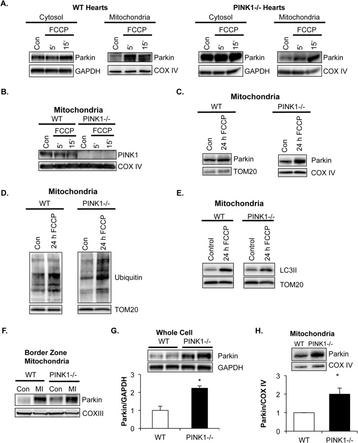
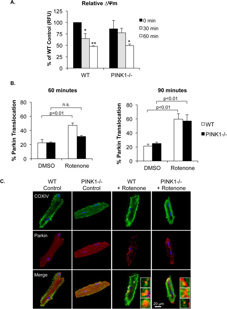
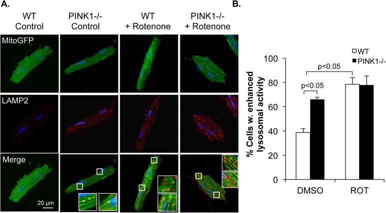
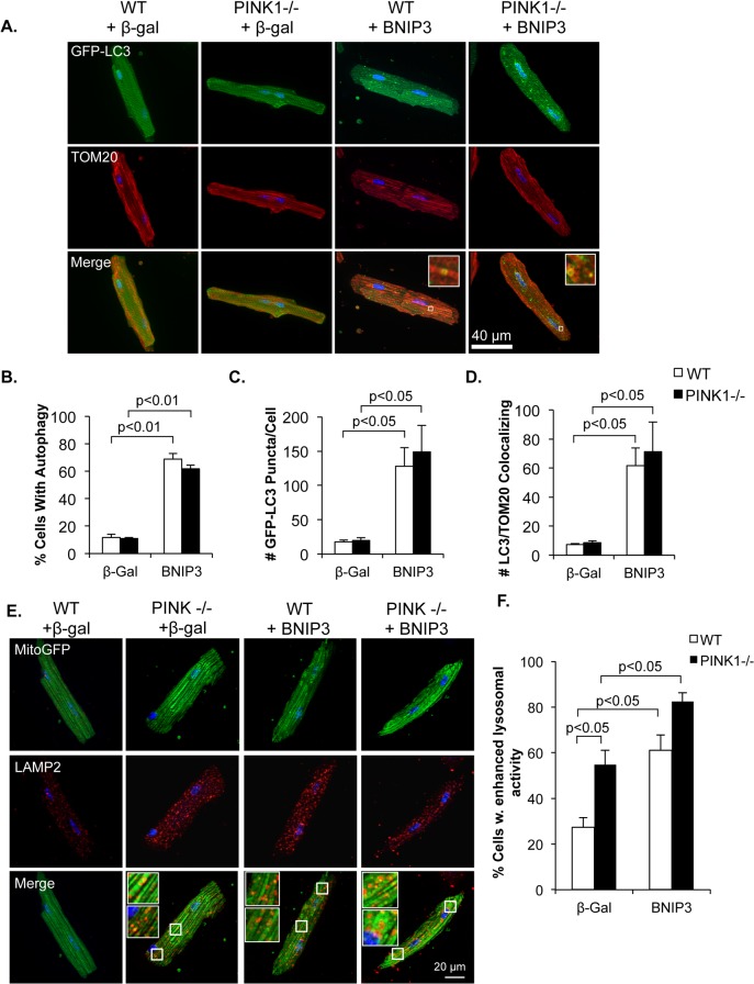
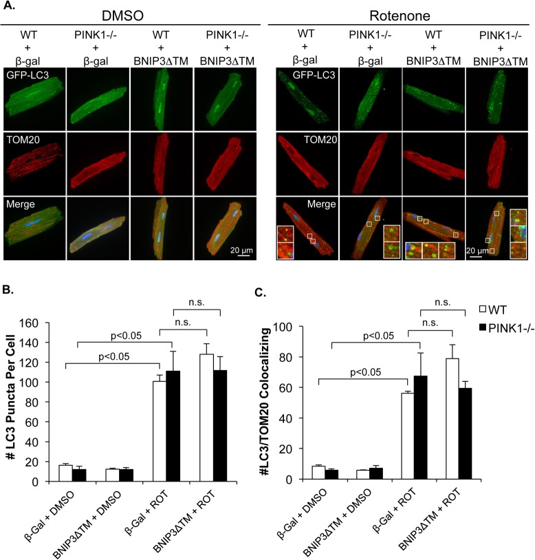
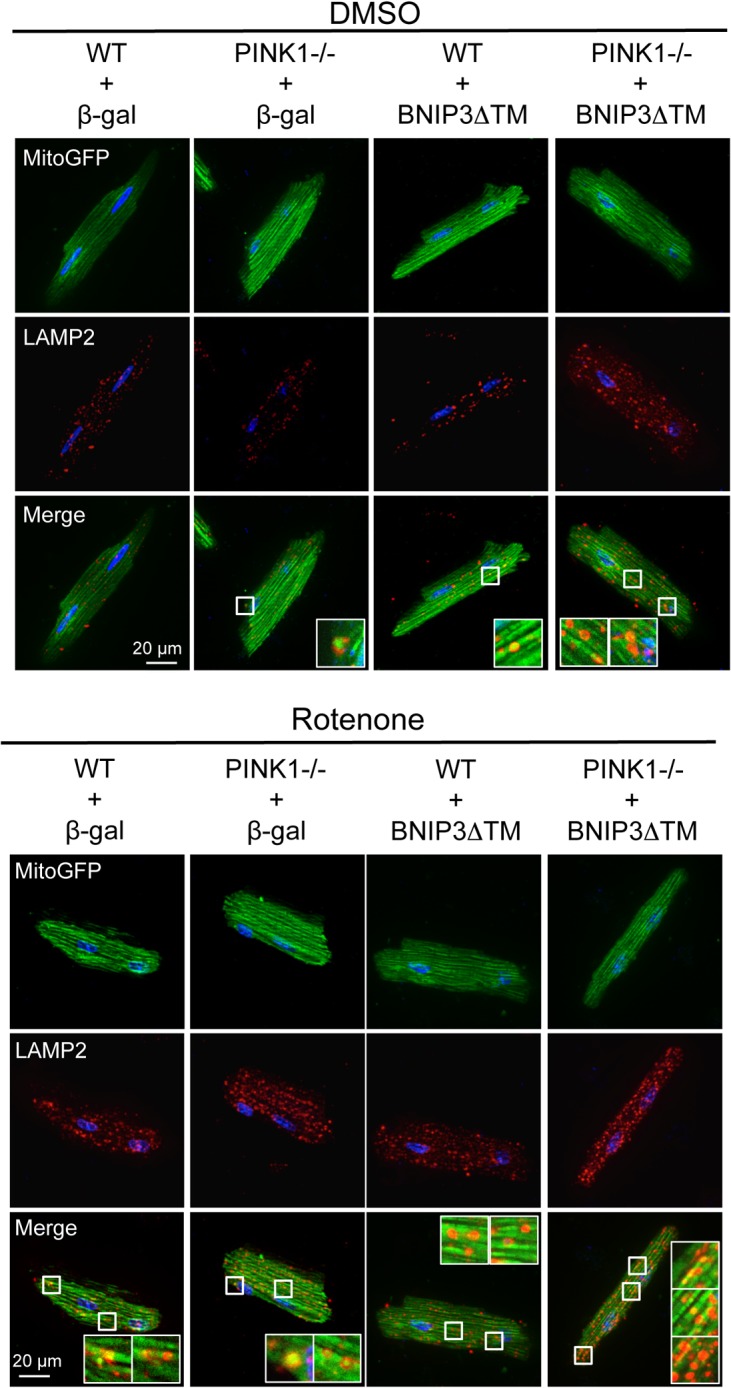
Myocyte function and survival relies on the maintenance of a healthy population of mitochondria. The PINK1/Parkin pathway plays an important role in clearing defective mitochondria via autophagy in cells. However, how the PINK1/Parkin pathway regulates mitochondrial quality control and whether it coordinates with other mitophagy pathways are still unclear. Therefore, the objective of this study was to investigate the effect of PINK1-deficiency on mitochondrial quality control in myocytes. Using PINK1-deficient (PINK1-/-) mice, we found that Parkin is recruited to damaged cardiac mitochondria in hearts after treatment with the mitochondrial uncoupler FCCP or after a myocardial infarction even in the absence of PINK1. Parkin recruitment to depolarized mitochondria correlates with increased ubiquitination of mitochondrial proteins and activation of mitophagy in PINK1-/- myocytes. In addition, induction of mitophagy by the atypical BH3-only protein BNIP3 is unaffected by lack of PINK1. Overall, these data suggest that Parkin recruitment to depolarized cardiac mitochondria and subsequent activation of mitophagy is independent of PINK1. Moreover, alternative mechanisms of Parkin activation and pathways of mitophagy remain functional in PINK1-/- myocytes and could compensate for the PINK1 deficiency.


Conflict of interest statement

Competing Interests: The authors have declared that no competing interests exist.

Figures

Fig 1
Fig 1. Translocation of Parkin to mitochondria and activation of mitophagy occur independently of PINK1.
(A) Perfusion of WT and PINK1-/- hearts with the mitochondrial uncoupler FCCP (100 nM) led to accumulation of Parkin in the mitochondrial fraction within 15 minutes (15’). (B) Perfusion of WT hearts with FCCP for up to 15 minutes did not result in accumulation of PINK1 in the mitochondrial fraction. PINK1-/- perfused heart samples shown for comparison. In vivo FCCP treatment for 24 h led to (C) translocation of Parkin to cardiac mitochondria, (D) increased ubiquitination of cardiac mitochondrial proteins, and (E) increased LC3II association with mitochondria. (F) Analysis of mitochondria isolated from the infarct border zone four hours after in vivo myocardial infarction (MI). Parkin levels increased in the mitochondrial fraction in both WT and PINK1-/- hearts after MI. Representative western blots and densitometry data show significantly elevated Parkin protein levels in the heart (G) and at the mitochondria (H) in 3 month old PINK1-/- mice. Mean ± SEM, n = 3, *p<0.05 vs. WT.
Fig 2
Fig 2. Rotenone induces delayed Parkin translocation to mitochondria in PINK1-/- myocytes.
(A) The mitochondrial complex I inhibitor rotenone (40 μM) significantly reduced mitochondrial membrane potential by 1 hour of treatment in WT and PINK1-/- cardiac myocytes (n = 3). (B) Quantitation of percentage of cells with Parkin translocation to mitochondria after 60 and 90 min of rotenone treatment. (C) Representative images. Mean ± SEM, n = 3, *p<0.05, **p<0.01 vs. 0 min. n.s. = not significant
Fig 3
Fig 3. Rotenone induces activation of autophagy and mitophagy in WT and PINK1-/- adult mouse myocytes.
(A) Quantitation of autophagy activation after 60 and 90 min rotenone (40 μM) treatment in WT and PINK1-/- myocytes. (B) Representative fluorescent images of WT and PINK1-/- myocytes overexpressing GFP-LC3 and stained with anti-TOM20 to label mitochondria. Co-localization between GFP-LC3 and TOM20 indicate activation of mitophagy. (C) Quantitation of the number of GFP-LC3 puncta in cells. (D) Quantitation of the number of GFP-LC3 autophagosomes co-localizing with TOM20-labeled mitochondria. (E) Transmission electron microscopy of isolated adult mouse myocytes after treatment with DMSO or 40 μM rotenone. Autophagosomes containing mitochondria were identified in both WT and PINK1-/- cardiac myocytes after rotenone treatment. Mean ± SEM, n = 3. n.s. = not significant
Fig 4
Fig 4. Autophagic flux is intact in rotenone treated PINK1-/- myocytes.
(A) Representative images of WT and PINK1-/- myocytes overexpressing MitoGFP and stained with anti-LAMP2 after 90 min of rotenone treatment. (B) Quantitation of WT and PINK1-/- myocytes with enhanced lysosomal activity. Mean ± SEM, n = 3.
Fig 5
Fig 5. BNIP3 induces mitophagy in WT and PINK1-/- myocytes.
(A) Representative fluorescent images of WT and PINK1-/- adult mouse cardiac myocytes overexpressing GFP-LC3 and either β-gal or BNIP3. (B) The percentage of cells with activated autophagy significantly increased in both WT and PINK1-/- cells in response to BNIP3 overexpression. (C) Quantitation of GFP-LC3 puncta in adult mouse cardiac myocytes show no difference in autophagosome number in PINK1-/- cells compared to WT. (D) Quantitation of GFP-LC3 puncta colocalization with mitochondrial TOM20 show increased mitophagy in both WT and PINK1-/- myocytes in response to BNIP3. (E) Representative images of LAMP2 stained WT and PINK1-/- myocytes overexpressing β-gal or BNIP3. (F) Quantitation of WT and PINK1-/- myocytes with enhanced lysosomal activity. Mean ± SEM, n = 3–4.
Fig 6
Fig 6. The dominant negative BNIP3ΔTM has no affect rotenone-mediated mitophagy.
(A) Representative fluorescent images of WT and PINK1-/- adult mouse cardiac myocytes overexpressing β-gal or BNIP3ΔTM plus GFP-LC3. (B) Quantitation of the number of autophagosomes in WT and PINK1-/- myocytes. (C) Quantitation of GFP-LC3 puncta colocalization with mitochondrial TOM20. BNIP3ΔTM did not reduce rotenone-mediated mitophagy in either WT or PINK1-/- myocytes. Mean ± SEM, n = 3. n.s. = not significant
Fig 7
Fig 7. Bnip3ΔTM does not affect rotenone-stimulated autophagic flux.
Representative images of LAMP2 stained WT and PINK1-/- myocytes overexpressing β-gal or BNIP3ΔTM plus MitoGFP after rotenone treatment (90 min).

Similar articles

Cited by

References

    1. Rizzuto R, De Stefani D, Raffaello A, Mammucari C. Mitochondria as sensors and regulators of calcium signalling. Nat Rev Mol Cell Biol. 2012;13(9):566–78. 10.1038/nrm3412 . - DOI - PubMed
    1. Tait SW, Green DR. Mitochondria and cell death: outer membrane permeabilization and beyond. Nat Rev Mol Cell Biol. 2010;11(9):621–32. 10.1038/nrm2952 . - DOI - PubMed
    1. Hammerling BC, Gustafsson AB. Mitochondrial quality control in the myocardium: cooperation between protein degradation and mitophagy. J Mol Cell Cardiol. 2014;75:122–30. 10.1016/j.yjmcc.2014.07.013 - DOI - PMC - PubMed
    1. Soubannier V, McLelland GL, Zunino R, Braschi E, Rippstein P, Fon EA, et al. A vesicular transport pathway shuttles cargo from mitochondria to lysosomes. Curr Biol. 2012;22(2):135–41. 10.1016/j.cub.2011.11.057 . - DOI - PubMed
    1. Kubli DA, Gustafsson AB. Mitochondria and mitophagy: the yin and yang of cell death control. Circ Res. 2012;111(9):1208–21. Epub 2012/10/16. doi: 111/9/1208 [pii]10.1161/CIRCRESAHA.112.265819 - DOI - PMC - PubMed

Publication types

LinkOut - more resources