

Breast Tissue Composition and Immunophenotype and Its Relationship with Mammographic Density in Women at High Risk of Breast Cancer
- PMID: 26110820
- PMCID: PMC4481506
- DOI: 10.1371/journal.pone.0128861
Breast Tissue Composition and Immunophenotype and Its Relationship with Mammographic Density in Women at High Risk of Breast Cancer
Abstract
Aim: To investigate the cellular and immunophenotypic basis of mammographic density in women at high risk of breast cancer.
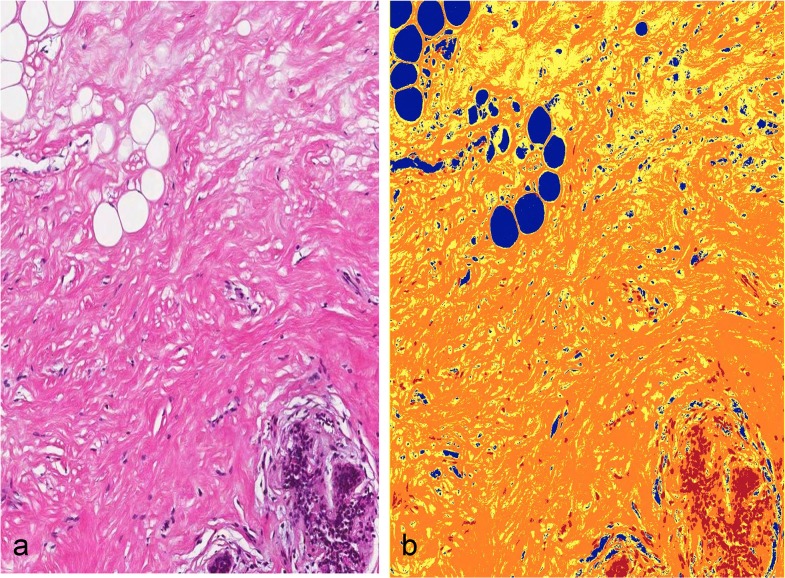
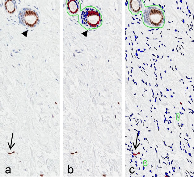
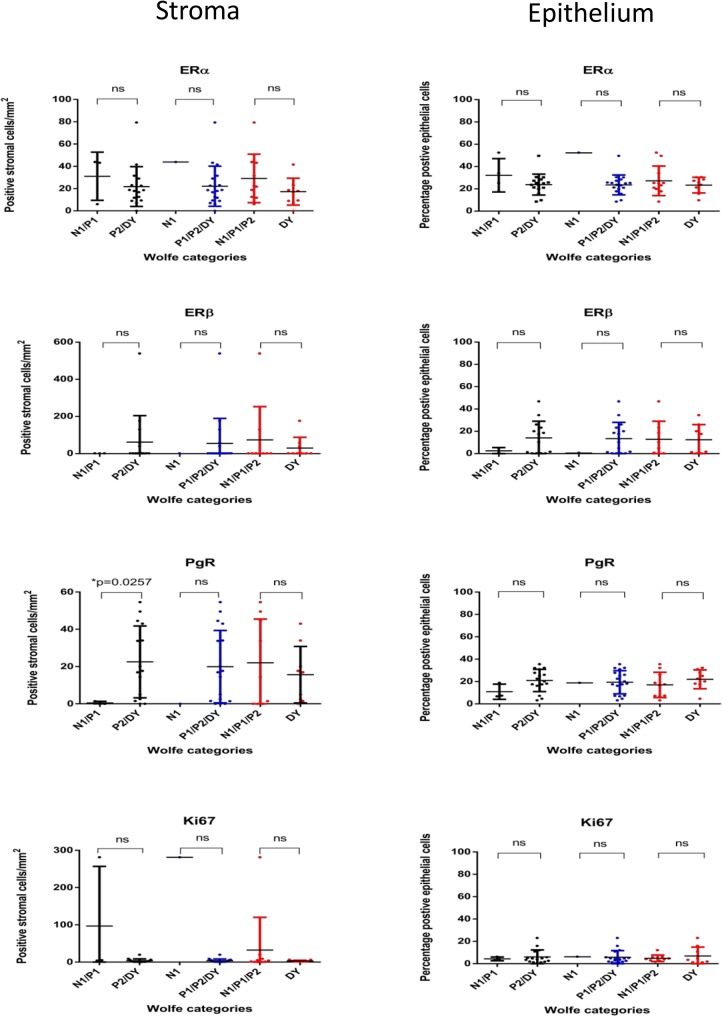
Methods: Mammograms and targeted breast biopsies were accrued from 24 women at high risk of breast cancer. Mammographic density was classified into Wolfe categories and ranked by increasing density. The histological composition and immunophenotypic profile were quantified from digitized haematoxylin and eosin-stained and immunohistochemically-stained (ERα, ERβ, PgR, HER2, Ki-67, and CD31) slides and correlated to mammographic density.
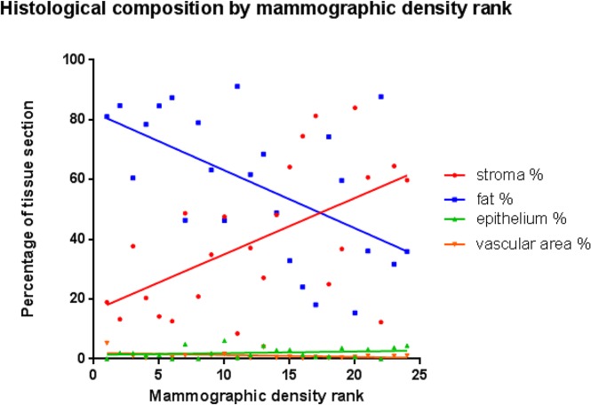
Results: Increasing mammographic density was significantly correlated with increased fibrous stroma proportion (rs (22) = 0.5226, p = 0.0088) and significantly inversely associated with adipose tissue proportion (rs (22) = -0.5409, p = 0.0064). Contrary to previous reports, stromal expression of ERα was common (19/20 cases, 95%). There was significantly higher stromal PgR expression in mammographically-dense breasts (p=0.026).
Conclusions: The proportion of stroma and fat underlies mammographic density in women at high risk of breast cancer. Increased expression of PgR in the stroma of mammographically dense breasts and frequent and unexpected presence of stromal ERα expression raises the possibility that hormone receptor expression in breast stroma may have a role in mediating the effects of exogenous hormonal therapy on mammographic density.
Conflict of interest statement
Figures
Similar articles
-
High mammographic density is associated with an increase in stromal collagen and immune cells within the mammary epithelium.Breast Cancer Res. 2015 Jun 4;17(1):79. doi: 10.1186/s13058-015-0592-1. Breast Cancer Res. 2015. PMID: 26040322 Free PMC article.
-
Amount of stroma is associated with mammographic density and stromal expression of oestrogen receptor in normal breast tissues.Breast Cancer Res Treat. 2016 Jul;158(2):253-61. doi: 10.1007/s10549-016-3877-x. Epub 2016 Jun 27. Breast Cancer Res Treat. 2016. PMID: 27349429
-
Mammographic density is related to stroma and stromal proteoglycan expression.Breast Cancer Res. 2003;5(5):R129-35. doi: 10.1186/bcr622. Epub 2003 Jul 23. Breast Cancer Res. 2003. PMID: 12927043 Free PMC article.
-
Does mammographic density reflect the expression of breast cancer markers?Climacteric. 2013 Aug;16(4):407-16. doi: 10.3109/13697137.2013.798271. Epub 2013 Jun 4. Climacteric. 2013. PMID: 23617937 Review.
-
Stromal characteristics may hold the key to mammographic density: the evidence to date.Oncotarget. 2016 May 24;7(21):31550-62. doi: 10.18632/oncotarget.6912. Oncotarget. 2016. PMID: 26784251 Free PMC article. Review.
Cited by
-
Breast density is strongly associated with multiparametric magnetic resonance imaging biomarkers and pro-tumorigenic proteins in situ.Br J Cancer. 2022 Nov;127(11):2025-2033. doi: 10.1038/s41416-022-01976-3. Epub 2022 Sep 22. Br J Cancer. 2022. PMID: 36138072 Free PMC article.
-
Mammographic density by time and breast: a retrospective cohort study from BreastScreen Norway.Breast Cancer Res. 2025 May 16;27(1):83. doi: 10.1186/s13058-025-02037-2. Breast Cancer Res. 2025. PMID: 40380195 Free PMC article.
-
Stiff stroma increases breast cancer risk by inducing the oncogene ZNF217.J Clin Invest. 2020 Nov 2;130(11):5721-5737. doi: 10.1172/JCI129249. J Clin Invest. 2020. PMID: 32721948 Free PMC article. Clinical Trial.
-
Equal Pro-inflammatory Profiles of CCLs, CXCLs, and Matrix Metalloproteinases in the Extracellular Microenvironment In Vivo in Human Dense Breast Tissue and Breast Cancer.Front Immunol. 2018 Jan 16;8:1994. doi: 10.3389/fimmu.2017.01994. eCollection 2017. Front Immunol. 2018. PMID: 29387062 Free PMC article.
-
Mammographic density: a potential monitoring biomarker for adjuvant and preventative breast cancer endocrine therapies.Oncotarget. 2017 Jan 17;8(3):5578-5591. doi: 10.18632/oncotarget.13484. Oncotarget. 2017. PMID: 27894075 Free PMC article. Review.
References
-
- Boyd NF, Dite GS, Stone J, Gunasekara A, English DR, McCredie MR, et al. (2002) Heritability of mammographic density, a risk factor for breast cancer. N Engl J Med 347: 886–894. - PubMed
-
- Li T, Sun L, Miller N, Nicklee T, Woo J, Hulse-Smith L, et al. (2005) The association of measured breast tissue characteristics with mammographic density and other risk factors for breast cancer. Cancer Epidemiol Biomarkers Prev 14: 343–349. - PubMed
Publication types
MeSH terms
Substances
LinkOut - more resources
Full Text Sources
Other Literature Sources
Medical
Research Materials
Miscellaneous
