

Tetraarsenictetrasulfide and Arsenic Trioxide Exert Synergistic Effects on Induction of Apoptosis and Differentiation in Acute Promyelocytic Leukemia Cells
- PMID: 26110921
- PMCID: PMC4481354
- DOI: 10.1371/journal.pone.0130343
Tetraarsenictetrasulfide and Arsenic Trioxide Exert Synergistic Effects on Induction of Apoptosis and Differentiation in Acute Promyelocytic Leukemia Cells
Abstract
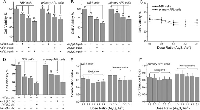
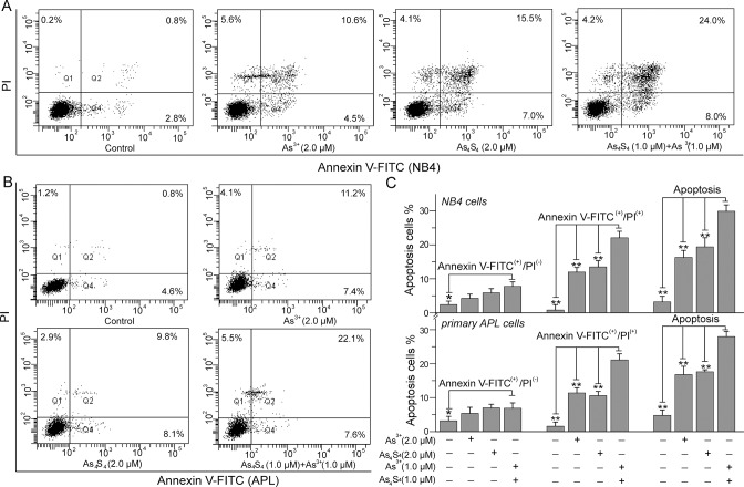
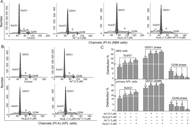
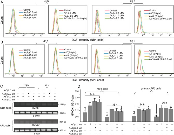
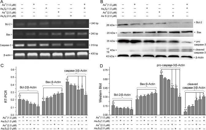
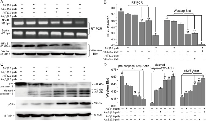
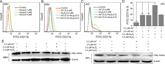
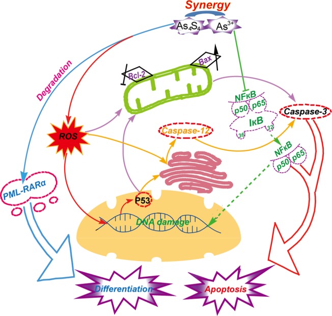
Since arsenic trioxide (As3+) has been successfully used in the treatment of acute promyelocytic leukemia (APL), its adverse effects on patients have been problematic and required a solution. Considering the good therapeutic potency and low toxicity of tetraarsenictetrasulfide (As4S4) in the treatment of APL, we investigated the effects of combining As4S4 and As3+ on the apoptosis and differentiation of NB4 and primary APL cells. As4S4, acting similarly to As3+, arrested the G1/S transition, induced the accumulation of cellular reactive oxygen species, and promoted apoptosis. Additionally, low concentrations of As4S4 (0.1-0.4 μM) induced differentiation of NB4 and primary APL cells. Compared with the As4S4- or As3+-treated groups, the combination of As4S4 and As3+ obviously promoted apoptosis and differentiation of NB4 and primary APL cells. Mechanistic studies suggested that As4S4 acted synergistically with As3+ to down-regulate Bcl-2 and nuclear factor-κB expression, up-regulate Bax and p53 expression, and induce activation of caspase-12 and caspase-3. Moreover, the combination of low concentrations of As4S4 and As3+ enhanced degradation of the promyelocytic leukemia-retinoic acid receptor α oncoprotein. In summary, As4S4 and As3+ synergistically induce the apoptosis and differentiation of NB4 and primary APL cells.
Conflict of interest statement
Figures
Similar articles
-
Arsenic sulfide promotes apoptosis in retinoid acid resistant human acute promyelocytic leukemic NB4-R1 cells through downregulation of SET protein.PLoS One. 2014 Jan 13;9(1):e83184. doi: 10.1371/journal.pone.0083184. eCollection 2014. PLoS One. 2014. PMID: 24454695 Free PMC article.
-
Src family kinase inhibitor PP2 enhances differentiation of acute promyelocytic leukemia cell line induced by combination of all-trans-retinoic acid and arsenic trioxide.Leuk Res. 2014 Aug;38(8):977-82. doi: 10.1016/j.leukres.2014.05.019. Epub 2014 Jun 4. Leuk Res. 2014. PMID: 24953245
-
Sodium selenite induces apoptosis in acute promyelocytic leukemia-derived NB4 cells by a caspase-3-dependent mechanism and a redox pathway different from that of arsenic trioxide.Ann Hematol. 2004 Dec;83(12):751-8. doi: 10.1007/s00277-004-0920-5. Epub 2004 Oct 6. Ann Hematol. 2004. PMID: 15480664
-
Understanding the molecular pathogenesis of acute promyelocytic leukemia.Best Pract Res Clin Haematol. 2014 Mar;27(1):3-9. doi: 10.1016/j.beha.2014.04.006. Epub 2014 Apr 13. Best Pract Res Clin Haematol. 2014. PMID: 24907012 Review.
-
The use of arsenic trioxide (As2O3) in the treatment of acute promyelocytic leukemia.J Biol Regul Homeost Agents. 1999 Oct-Dec;13(4):195-200. J Biol Regul Homeost Agents. 1999. PMID: 10703942 Review.
Cited by
-
Solanine induced apoptosis and increased chemosensitivity to Adriamycin in T-cell acute lymphoblastic leukemia cells.Oncol Lett. 2018 May;15(5):7383-7388. doi: 10.3892/ol.2018.8229. Epub 2018 Mar 12. Oncol Lett. 2018. PMID: 29731890 Free PMC article.
-
BKM120 sensitizes BRCA-proficient triple negative breast cancer cells to olaparib through regulating FOXM1 and Exo1 expression.Sci Rep. 2021 Feb 26;11(1):4774. doi: 10.1038/s41598-021-82990-y. Sci Rep. 2021. PMID: 33637776 Free PMC article.
-
As4S4 Exhibits Good Killing Effect on Multiple Myeloma Cells Via Repressing SOCS1 Methylation-Mediated JAK2/STAT3 Signaling Pathway.Technol Cancer Res Treat. 2019 Jan-Dec;18:1533033819896806. doi: 10.1177/1533033819896806. Technol Cancer Res Treat. 2019. PMID: 31868118 Free PMC article.
-
Prohibitin 1 inhibits cell proliferation and induces apoptosis via the p53-mediated mitochondrial pathway in vitro.World J Gastrointest Oncol. 2024 Feb 15;16(2):398-413. doi: 10.4251/wjgo.v16.i2.398. World J Gastrointest Oncol. 2024. PMID: 38425403 Free PMC article.
-
Major apoptotic mechanisms and genes involved in apoptosis.Tumour Biol. 2016 Jul;37(7):8471-86. doi: 10.1007/s13277-016-5035-9. Epub 2016 Apr 9. Tumour Biol. 2016. PMID: 27059734 Review.
References
-
- Degos L (2003) The history of acute promyelocytic leukaemia. Brit J Haematol 122: 539–553. - PubMed
-
- De Thé H, Chomienne C, Lanotte M, Degos L, Dejean A (1990) The t(15;17) translocation of acute promyelocytic leukaemia fuses the retinoic acid receptor alpha gene to a novel transcribed locus. Nature 347: 558–561. - PubMed
-
- Huang ME, Ye YC, Chen SR, Chai JR, Lu JX, Zhoa L, et al. (1988) Use of all-trans retinoic acid in the treatment of acute promyelocytic leukemia. Blood 72: 567–572. - PubMed
Publication types
MeSH terms
Substances
LinkOut - more resources
Full Text Sources
Other Literature Sources
Research Materials
Miscellaneous
