Possible Genetic Origin of Limb-Body Wall Complex
- PMID: 26111189
- PMCID: PMC4673583
- DOI: 10.3109/15513815.2015.1055021
Possible Genetic Origin of Limb-Body Wall Complex
Abstract
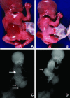
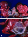
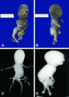
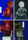
Limb body wall complex (LBWC) is characterized by multiple severe congenital malformations including an abdominal and/or thoracic wall defect covered by amnion, a short or absent umbilical cord with the placenta almost attached to the anterior fetal wall, intestinal malrotation, scoliosis, and lower extremity anomalies. There is no consensus about the etiology of LBWC and many cases with abnormal facial cleft do not meet the requirements for the true complex. We describe a series of four patients with LBWC and other malformations in an attempt to explain their etiology. There are several reports of fetuses with LBWC and absent gallbladder and one of our patients also had polysplenia. Absent gallbladder and polysplenia are associated with laterality genes including HOX, bFGF, transforming growth factor beta/activins/BMP4, WNT 1-8, and SHH. We postulate that this severe malformation may be due to abnormal genes involved in laterality and caudal development.
Keywords: absent gallbladder; body stalk anomaly; genes; limb body wall complex (lbwc); polysplenia; short umbilical cord.
Figures


Similar articles
-
Limb-body wall complex: 4 new cases illustrating the importance of examining placenta and umbilical cord.Pathol Res Pract. 2000;196(11):783-90. doi: 10.1016/S0344-0338(00)80114-4. Pathol Res Pract. 2000. PMID: 11186176
-
A case of limb-body wall complex diagnosed in utero.J Emerg Med. 2003 Jul;25(1):104-6. doi: 10.1016/s0736-4679(03)00122-7. J Emerg Med. 2003. PMID: 12865119 No abstract available.
-
Patterns of severe abdominal wall defects: insights into pathogenesis, delineation, and nomenclature.Birth Defects Res A Clin Mol Teratol. 2007 Mar;79(3):211-20. doi: 10.1002/bdra.20339. Birth Defects Res A Clin Mol Teratol. 2007. PMID: 17183587
-
Fetal abdominal wall defects.Best Pract Res Clin Obstet Gynaecol. 2014 Apr;28(3):391-402. doi: 10.1016/j.bpobgyn.2013.10.003. Epub 2013 Dec 3. Best Pract Res Clin Obstet Gynaecol. 2014. PMID: 24342556 Review.
-
[Antenatal diagnosis of limb body wall complex].J Gynecol Obstet Biol Reprod (Paris). 2000 Jun;29(4):385-91. J Gynecol Obstet Biol Reprod (Paris). 2000. PMID: 10844326 Review. French.
Cited by
-
Complex Body Wall Closure Defects in Seven Dog Fetuses: An Anatomic and CT Scan Study.Animals (Basel). 2025 Jul 10;15(14):2030. doi: 10.3390/ani15142030. Animals (Basel). 2025. PMID: 40723492 Free PMC article.
-
Insights into Congenital Body Stalk Anomaly Coupled with Placenta Accreta Conditions: A Case Report.Am J Case Rep. 2025 Apr 30;26:e946041. doi: 10.12659/AJCR.946041. Am J Case Rep. 2025. PMID: 40302190 Free PMC article.
-
BODY STALK ANOMALY: CLINICAL AND HISTOPATHOLOGIC FINDINGS OF THIS RARE ANOMALY IN A NIGERIAN NEWBORN.Ann Ib Postgrad Med. 2024 Apr 30;22(1):104-107. Ann Ib Postgrad Med. 2024. PMID: 38939879 Free PMC article.
-
Rare Presentation of Limb-Body Wall Complex in a Neonate: Case Report and Review of Literature.AJP Rep. 2022 Mar 7;12(1):e108-e112. doi: 10.1055/s-0042-1744215. eCollection 2022 Jan. AJP Rep. 2022. PMID: 35265395 Free PMC article.
-
Imaging findings in association with altered maternal alpha-fetoprotein levels during pregnancy.Abdom Radiol (NY). 2020 Oct;45(10):3239-3257. doi: 10.1007/s00261-020-02499-2. Abdom Radiol (NY). 2020. PMID: 32221672 Review.
References
-
- Van Allen MI, Curry C, Walden CE, Gallagher L, James F. Reynolds: Limb body wall complex: I. Pathogenesis. Am J Med Genet. 1987;28:529–548. - PubMed
-
- Miller ME. Structural defects as a consequence of early intrauterine constraint: Limb deficiency, polydactyly, and body wall defect. Semin Perinatol. 1983;7:274–277. - PubMed
-
- Bugge M. Body stalk anomaly in Denmark during 20 years (1970–1989) Am J Med Genet A. 2012;158A:1702. - PubMed
-
- Van Allen MI, Curry C, Walden CE, Gallagher L, James F. Reynolds: Limb body wall complex: II. Pathogenesis. Am J Med Genet. 1987;28:549–565. - PubMed
-
- Colpaert C, Bogers J, Hertveldt K, Loquet P, Dumon J, Willems P. Limb body wall complex: 4 new cases illustrating the importance of examining the cord and placenta. Pathol Res Pract. 2000;196:783–790. - PubMed
Publication types
MeSH terms
LinkOut - more resources
Full Text Sources
Other Literature Sources
