. 2015 Oct;14(5):887-95.
doi: 10.1111/acel.12368. Epub 2015 Jun 26.

Coenzyme Q10 restores oocyte mitochondrial function and fertility during reproductive aging

Affiliations

Coenzyme Q10 restores oocyte mitochondrial function and fertility during reproductive aging

Assaf Ben-Meir et al. Aging Cell. 2015 Oct.

Abstract

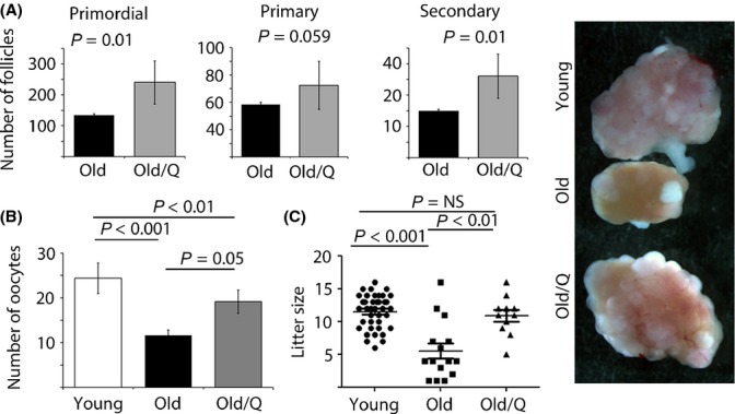
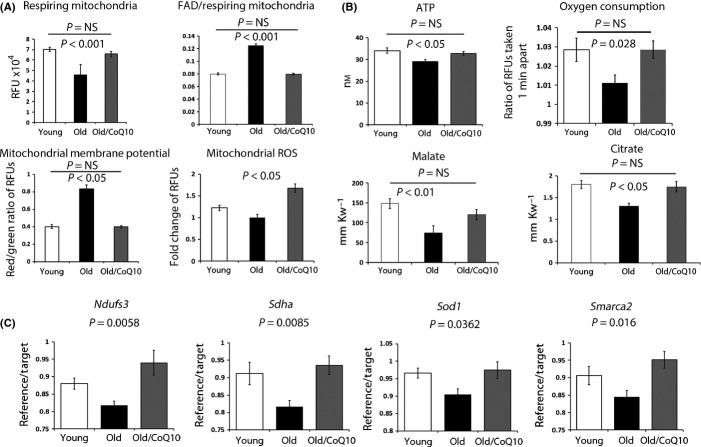
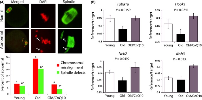
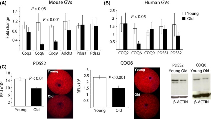
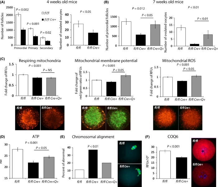
Female reproductive capacity declines dramatically in the fourth decade of life as a result of an age-related decrease in oocyte quality and quantity. The primary causes of reproductive aging and the molecular factors responsible for decreased oocyte quality remain elusive. Here, we show that aging of the female germ line is accompanied by mitochondrial dysfunction associated with decreased oxidative phosphorylation and reduced Adenosine tri-phosphate (ATP) level. Diminished expression of the enzymes responsible for CoQ production, Pdss2 and Coq6, was observed in oocytes of older females in both mouse and human. The age-related decline in oocyte quality and quantity could be reversed by the administration of CoQ10. Oocyte-specific disruption of Pdss2 recapitulated many of the mitochondrial and reproductive phenotypes observed in the old females including reduced ATP production and increased meiotic spindle abnormalities, resulting in infertility. Ovarian reserve in the oocyte-specific Pdss2-deficient animals was diminished, leading to premature ovarian failure which could be prevented by maternal dietary administration of CoQ10. We conclude that impaired mitochondrial performance created by suboptimal CoQ10 availability can drive age-associated oocyte deficits causing infertility.

Keywords: Mitochondria; anti-aging; fecundity; individual; molecular biology of aging; mouse models.


Figures

Fig 1
Fig 1
Impact of CoQ10 treatment on ovarian reserve and breeding performance in an aged mouse model. (A) Ovarian reserve was significantly higher in old vehicle mice treated with CoQ10 for a period of 15 weeks (n = 9/age and treatment) evidenced by significantly higher number of resting primordial and growing secondary follicles. Values represent average follicle numbers ± SEM. Images of stimulated ovaries from each group are shown on the right – magnification 50×. (B) Number of ovulated oocytes collected after hormonal stimulation of young (n = 20), old vehicle-treated (n = 16), and old CoQ10-treated dams (n = 16). Values represent average number of oocytes per female ± SEM. (C) Litter size in young (n = 39), old vehicle-treated (n = 15), and old CoQ10-treated mice (n = 11). The number of live pups born to dams in the 13th month of age was decreased, but normalized after CoQ10 supplementation. Each female produced only one litter during breeding trial. Scatter plot data are shown as mean per female ± SEM.
Fig 2
Fig 2
Improvement in mitochondrial function in oocytes of old vehicle mice after CoQ10 supplementation. (A) Oocytes from young, old vehicle and old CoQ10 mice were stained with MitoTracker Red, JC-1, and Mitosox or examined for green autofluorescence (FAD). The respiring mitochondrial pool (MitoTracker Red) was reduced in oocytes from old vehicle mice compared to young or old CoQ10, which were not different from each other. The ratio of oxidized FAD (FAD++)/MitoTracker Red increased in old oocytes. The mitochondrial membrane potential (MMP) increased in old oocytes, while ROS production (Mitosox) decreased. These aging effects were normalized by CoQ10 treatment. Values represent random fluorescence units (RFUs) per oocyte ± SEM. For all experiments, individual oocytes were used and groups contained n = 15–25 oocytes/age/treatment. (B) ATP (nm) and TCA cycle metabolites (millimole of substrate per kilogram wet weight per oocyte) were evaluated in individual oocytes from young (n = 7–15), old vehicle (n = 8–14), and old CoQ10 (n = 11–17). Oxygen consumption is expressed as a ratio of fluorescent signals obtained by a scan 1 min apart and reflects oxidative decay of the probe per oocyte. Data shown are mean ± SEM. (C) Expression levels of genes involved in mitochondrial function (Ndufs3, Sdha, and Sod1) and chromatin organization (Smarca2) in ovulated oocytes were reduced with aging and improved after treatment with CoQ10 (mean ± SEM). Each sample contained a pool of 3 oocytes (n = 4 young, n = 6 old vehicle and n = 5 old CoQ10 pools), and data are shown as the ratio of reference (actin)/target (studied) transcript.
Fig 3
Fig 3
CoQ10 rescues spindle defects in aging oocytes. (A) Percent of chromosomal or spindle misalignment in ovulated oocytes from young (n = 60), old (n = 73), and old CoQ10 (n = 51) mice. Oocytes were stained with antitubulin antibody (green) and DAPI (red). Representative images of normal spindle (barrel shaped) and chromosome alignment (toothbrush appearance) were considered normal. Arrows demonstrate detachment of chromosomes from spindle or misshaped spindle organization. Letters (a vs. b and a* vs. b*) are significantly different from each other (P < 0.05). (B) Expression of genes implicated in spindle formation/attachment and meiotic execution (Tuba1a, Hook1, Nek2, and Smarca2) in oocytes was reduced with aging and improved after treatment with CoQ10. Each sample contained a pool of 3 oocytes, and each age category was represented by n = 4 young, n = 6 old, and n = 5 old CoQ10 pools, and data are shown as the mean ratio of reference (actin)/target ± SEM.
Fig 4
Fig 4
Reduced expression of CoQ10 biosynthesis genes in oocytes with aging. (A) Fold change in mRNA level of pooled GVs (3 per sample; n = 6 young, n = 6 old) were normalized to young age. Transcripts encoding enzymes Pdss1 and Pdss2 did not change with aging, but the expression of the enzymes involved in modifying the hydroxybenzoate ring (Coq6 and Coq9) significantly decreased. (B) Fold change in mRNA level of CoQ10 synthesis genes in single human GV oocyte per patient (n = 8 patients <32 years old, n = 8 patients >39 years old females). Data are shown as mean ± SEM and are normalized to actin. (C) Immunocytochemistry of GV oocytes exposed to anti-PDSS2 and anti-COQ6 antibodies from young (n = 9) and old vehicle mice (n = 5). Values represent mean fluorescence units ± SEM. Western Blot of whole ovarian lysates from young (3 months old) and aged mice (12 months old) blotted with anti-PDSS2, anti-COQ6, and anti-actin antibodies.
Fig 5
Fig 5
Decreased ovarian reserve and oocyte quality in Pdss2 fl/fl Cre+ mice is rescued by CoQ10 supplementation. (A) The follicular count of ovaries (mean ± SEM) from 4-week-old vehicle mice revealed decreased ovarian reserve in Pdss2 fl/fl Cre+ (n = 8) compared to Pdss2 fl/fl Cre− mice (n = 5). Follicle loss is reflected also by the reduced ovulation rate (n = 8 Pdss2 fl/fl Cre+, n = 16 Pdss2 fl/fl Cre−). (B) Treatment with CoQ10 from birth till 7 weeks of age prevented loss of ovarian reserve (mean ± SEM) and improved the ovulation rate triggered by Pdss2 deficiency. (C) Oocytes from Pdss2 fl/fl Cre+ exhibit mitochondrial dysfunction with decreased respiring mitochondrial pool, reduced mitochondrial membrane potential (MMP), and decreased ROS production. Similar to aging, MMP and mitochondrial ROS levels were significantly improved under CoQ10 supplementation. (D) ATP output per oocyte improved by CoQ10 administration in Pdss2 fl/fl Cre+ females. All data shown are mean ± SEM obtained from 20–40 oocytes. (E) Chromosomal misalignment was significantly more frequent in oocytes from Pdss2 fl/fl Cre+ (n = 62) compared to Pdss2 fl/fl Cre− (n = 118), and this was corrected by CoQ10 administration (n = 56). (F) Anti-COQ6 protein level in growing GV oocytes. Pdss2 fl/fl Cre+ oocytes present with significantly reduced levels of CoQ6 protein (n = 31 Pdss2 fl/fl Cre−, n = 44 Pdss2 fl/fl Cre+).

References

    1. Battaglia DE, Goodwin P, Klein NA, Soules MR. Influence of maternal age on meiotic spindle assembly in oocytes from naturally cycling women. Hum. Reprod. 1996;11:2217–2222. - PubMed
    1. Bentov Y, Yavorska T, Esfandiari N, Jurisicova A, Casper RF. The contribution of mitochondrial function to reproductive aging. J. Assist. Reprod. Genet. 2011;28:773–783. - PMC - PubMed
    1. Chi MM, Hoehn A, Moley KH. Metabolic changes in the glucose-induced apoptotic blastocyst suggest alterations in mitochondrial physiology. Am. J. Physiol. Endocrinol. Metab. 2002;283:E226–E232. - PubMed
    1. Chiang T, Schultz RM, Lampson MA. Age-dependent susceptibility of chromosome cohesion to premature separase activation in mouse oocytes. Biol. Reprod. 2011;85:1279–1283. - PMC - PubMed
    1. Crane FL. Biochemical functions of coenzyme Q10. J. Am. Coll. Nutr. 2001;20:591–598. - PubMed

Publication types