. 2015 Aug;35(8):1787-97.
doi: 10.1161/ATVBAHA.115.305609. Epub 2015 Jun 25.

Foamy monocytes form early and contribute to nascent atherosclerosis in mice with hypercholesterolemia

Affiliations

Foamy monocytes form early and contribute to nascent atherosclerosis in mice with hypercholesterolemia

Lu Xu et al. Arterioscler Thromb Vasc Biol. 2015 Aug.

Abstract

Objective: To examine infiltration of blood foamy monocytes, containing intracellular lipid droplets, into early atherosclerotic lesions and its contribution to development of nascent atherosclerosis.

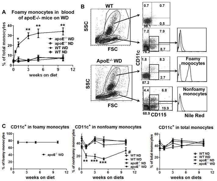
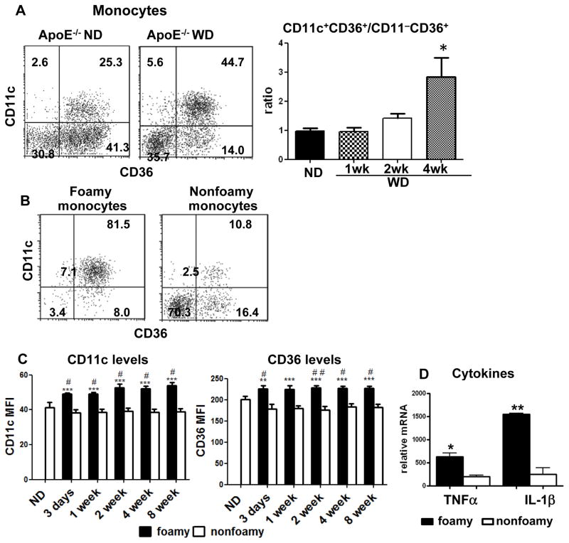
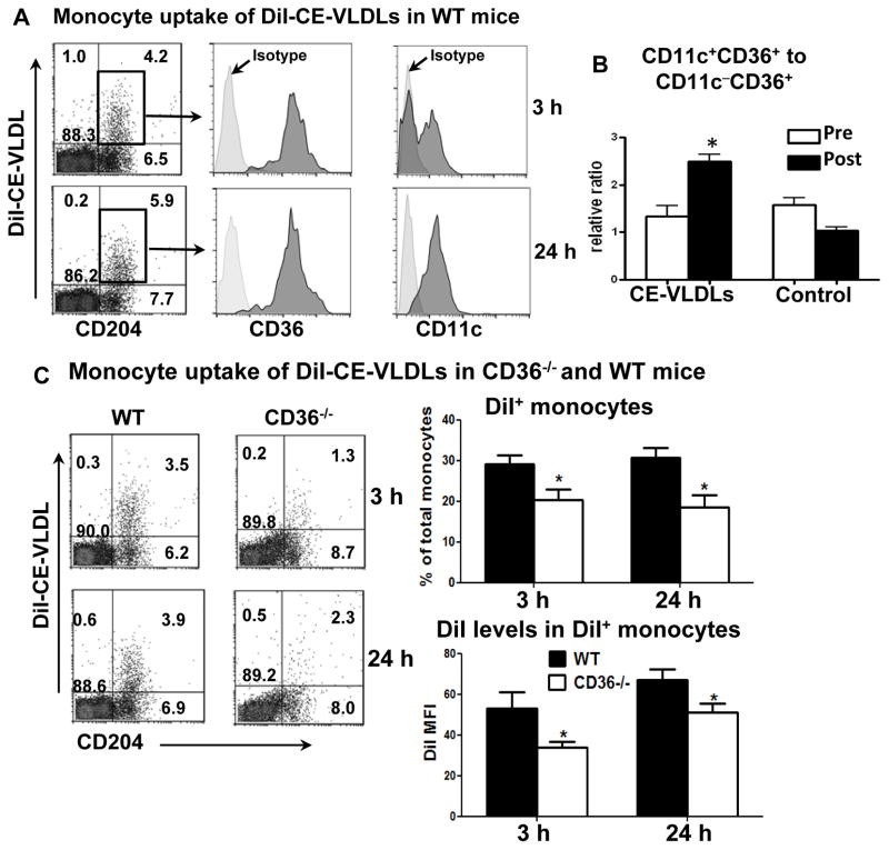
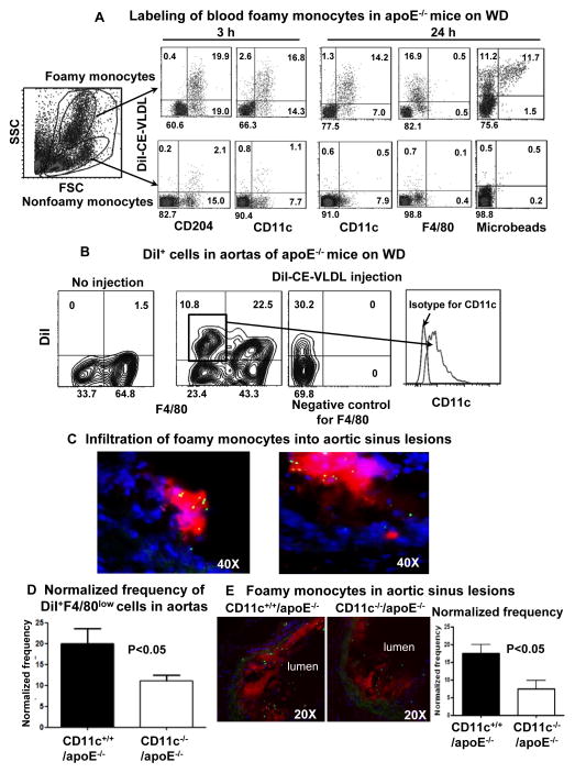
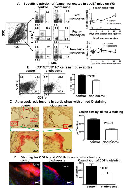
Approach and results: In apoE(-/-) mice fed Western high-fat diet (WD), >10% of circulating monocytes became foamy monocytes at 3 days on WD and >20% of monocytes at 1 week. Foamy monocytes also formed early in blood of Ldlr(-/-)Apobec1(-/-) (LDb) mice on WD. Based on CD11c and CD36, mouse monocytes were categorized as CD11c(-)CD36(-), CD11c(-)CD36(+), and CD11c(+)CD36(+). The majority of foamy monocytes were CD11c(+)CD36(+), whereas most nonfoamy monocytes were CD11c(-)CD36(-) or CD11c(-)CD36(+) in apoE(-/-) mice on WD. In wild-type mice, CD11c(+)CD36(+) and CD11c(-)CD36(+), but few CD11c(-)CD36(-), monocytes took up cholesteryl ester-rich very low-density lipoproteins (CE-VLDLs) isolated from apoE(-/-) mice on WD, and CE-VLDL uptake accelerated CD11c(-)CD36(+) to CD11c(+)CD36(+) monocyte differentiation. Ablation of CD36 decreased monocyte uptake of CE-VLDLs. Intravenous injection of DiI-CE-VLDLs in apoE(-/-) mice on WD specifically labeled CD11c(+)CD36(+) foamy monocytes, which infiltrated into nascent atherosclerotic lesions and became CD11c(+) cells that were selectively localized in atherosclerotic lesions. CD11c deficiency reduced foamy monocyte infiltration into atherosclerotic lesions. Specific and consistent depletion of foamy monocytes (for 3 weeks) by daily intravenous injections of low-dose clodrosome reduced development of nascent atherosclerosis.

Conclusions: Foamy monocytes, which form early in blood of mice with hypercholesterolemia, infiltrate into early atherosclerotic lesions in a CD11c-dependent manner and play crucial roles in nascent atherosclerosis development.

Keywords: atherosclerosis; diet, high-fat; inflammation; lipoproteins; monocytes.


Figures

Figure 1
Figure 1. Early appearance of foamy monocytes in blood of apoE−/− mice on western high-fat diet (WD)
A, Percentages of foamy monocytes in total monocytes in blood of apoE−/− mice at various time points (from 3 days to 10 weeks) after starting WD analyzed by flow cytometry. n=6–7 mice/group. B, Representative flow cytometric examples of monocytes in WT or apoE−/− mice on WD (for 3 days) from more than 5 independent experiments with 3–7 samples/group in each experiment. C, Percentages of CD11c+ monocytes in foamy, nonfoamy and total monocytes in blood of apoE−/− and WT mice on WD or normal diet (ND). n=6–7 mice/group. *P<0.05 versus WT WD and P<0.01 versus WT ND and apoE−/− ND; **P<0.01, ***P<0.001 versus WT ND, WT WD and apoE−/− ND; #P<0.05 versus WT ND and WT WD.
Figure 2
Figure 2. Three major monocyte subsets in mouse blood
A, A representative sample of monocyte subsets in blood of a WT mouse showing three major subsets based on CD11c and CD36, and their relationship to Ly-6C expression, from 3 independent experiments with 3 or 4 samples in each experiment. B, A representative example of monocyte reappearance and subset conversion in blood of WT mice after monocyte depletion by intravenous injection of clodrosome (0.3 ml/mouse), from 2 independent experiments with 4 samples in each experiment. C, A representative example of subset conversion of EdU-labeled monocytes in blood of WT mice after injection with EdU, from 2 independent experiments with 3 samples in each experiment.
Figure 3
Figure 3. Some characteristics of foamy monocytes in apoE−/− mice on WD
A, Representative examples of monocyte subsets (left panel) and relative ratios of CD11c+CD36+ to CD11cCD36+ monocytes (right panel) in apoE−/− mice on ND or WD. n=4–6 mice/group. B, Representative staining for CD11c and CD36 of foamy and nonfoamy monocytes in apoE−/− mice on WD (4 weeks) from more than 5 independent experiments with at least 4 samples in each experiment. C, Comparisons of CD11c and CD36 mean fluorescence intensity (MFI) levels on foamy and nonfoamy CD11c+CD36+ monocytes in apoE−/− mice on WD or ND. n=5–9 mice/group. D, mRNA levels of TNFα and IL-1β in foamy and nonfoamy monocytes from apoE−/− mice on WD. n=3 samples, each of which were pooled blood from 5 mice. *P<0.05, ***P<0.001 versus ND group (in panel A) or nonfoamy monocytes (in panels C and D); #P<0.05, ##P<0.01 versus ND group (in panel C).
Figure 4
Figure 4. Monocyte uptake of CE-VLDLs and subset conversion in WT and CD36−/− mice
A, Representative flow cytometric analyses of monocytes that took up DiI-CE-VLDLs (becoming DiI+), from more than 5 independent experiments with at least 3 samples in each experiment. A bolus of DiI-CE-VLDLs was intravenously injected into WT mice on ND. At 3 and 24 hours, blood was taken for flow cytometric analysis. Gated total leukocytes (for CD204) or DiI+ monocytes (for CD36 and CD11c) are presented. B, Relative ratios of CD11c+CD36+ to CD11cCD36+ monocytes in WT mice on ND after receiving repetitive daily injection of CE-VLDLs or PBS (control) for 3 days. n=4 mice/group. C, Representative flow cytometric analysis of monocyte uptake of DiI-CE-VLDLs, and quantification of DiI+ monocytes in total monocytes and DiI MFI levels on DiI+ monocytes, in WT and CD36−/− mice after receiving intravenous injection of a bolus of DiI-CE-VLDLs. n=11–12 mice/group. *P<0.05 versus preinjection (in panel B) or WT controls (in panel C).
Figure 5
Figure 5. Labeling of foamy monocytes and infiltration of foamy monocytes into nascent atherosclerotic lesions
A, Representative flow cytometric analysis samples showing specific labeling of foamy monocytes and phenotypes of labeled foamy monocytes in apoE−/− mice on WD (3 weeks) after intravenous injection of DiI-CE-VLDLs with or without fluorescent microbeads, from more than 5 independent experiments with at least 3 samples in each experiment. B, Representative flow cytometric analysis samples showing foamy monocytes (DiI+F4/80low) in atherosclerotic aortas with sustained expression of CD11c, from 4 independent experiments with at least 3 samples in each experiment. C, Representative histology of aortic sinus showing accumulation of microbead+ (green) cells, which were also DiI+ (red, left panel) and CD11c+ (red, right panel), in atherosclerotic lesions of apoE−/− mice on WD (3 weeks) after intravenous injection of fluorescent microbeads with (left panel) or without (right panel) DiI-CE-VLDLs. D, Normalized frequency of infiltrated foamy monocytes in aortas of CD11c−/−/apoE−/− and CD11c+/+/apoE−/− mice on WD (3 weeks). n=5–7 samples/group. E, Representative histology and quantification of foamy monocyte infiltration in aortic sinus lesions of CD11c−/−/apoE−/− and CD11c+/+/apoE−/− mice on WD (3 weeks). n=15 samples/group.
Figure 6
Figure 6. Effects of foamy monocyte depletion on development of nascent atherosclerosis
A, Specific depletion of foamy monocytes in apoE−/− mice on WD with daily injection of low-dose clodrosome (3 weeks). n=7 samples/group. B, CD11b+/CD11c+ cells in aortas of apoE−/− mice on WD with or without foamy monocyte depletion. n=5–7 samples/group. C, Quantification of atherosclerotic lesions, as indicated by oil red O staining, in aortic sinus of apoE−/− mice on WD with or without foamy monocyte depletion. n=5 mice/group. D, Representative staining for CD11c (red) and CD11b (green), and quantitation of CD11c staining in aortic sinus lesions of apoE−/− mice on WD with or without foamy monocyte depletion. n=5 mice/group.

References

    1. Libby P. Inflammation in atherosclerosis. Nature. 2002;420:868–874. - PubMed
    1. Koltsova EK, Garcia Z, Chodaczek G, Landau M, McArdle S, Scott SR, von Vietinghoff S, Galkina E, Miller YI, Acton ST, Ley K. Dynamic T cell-APC interactions sustain chronic inflammation in atherosclerosis. J Clin Invest. 2012;122:3114–3126. - PMC - PubMed
    1. Moore KJ, Sheedy FJ, Fisher EA. Macrophages in atherosclerosis: a dynamic balance. Nat Rev Immunol. 2013;13:709–721. - PMC - PubMed
    1. Woollard KJ, Geissmann F. Monocytes in atherosclerosis: subsets and functions. Nat Rev Cardiol. 2010;7:77–86. - PMC - PubMed
    1. Bilheimer DW, Goldstein JL, Grundy SM, Starzl TE, Brown MS. Liver transplantation to provide low-density-lipoprotein receptors and lower plasma cholesterol in a child with homozygous familial hypercholesterolemia. N Engl J Med. 1984;311:1658–1664. - PMC - PubMed

Publication types

MeSH terms