. 2015 Oct;23(10):1630-40.
doi: 10.1038/mt.2015.115. Epub 2015 Jun 26.

Viral Infection of Tumors Overcomes Resistance to PD-1-immunotherapy by Broadening Neoantigenome-directed T-cell Responses

Affiliations

Viral Infection of Tumors Overcomes Resistance to PD-1-immunotherapy by Broadening Neoantigenome-directed T-cell Responses

Norman Woller et al. Mol Ther. 2015 Oct.

Abstract

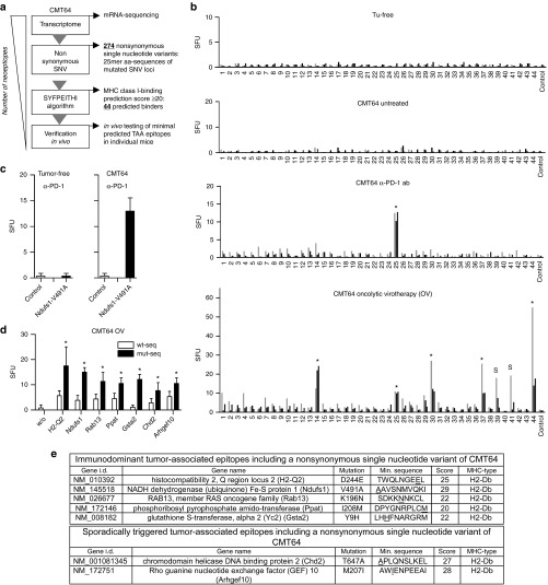
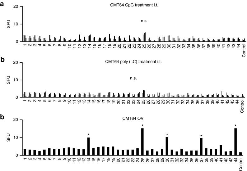
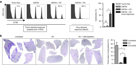
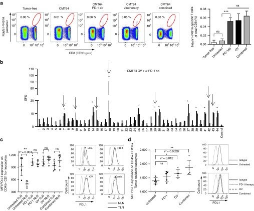
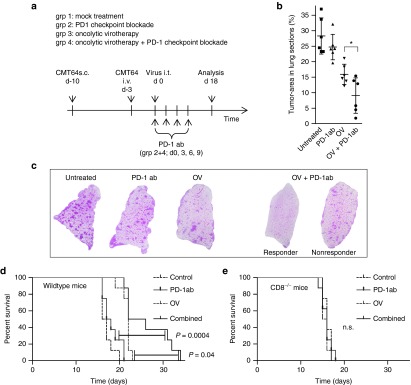
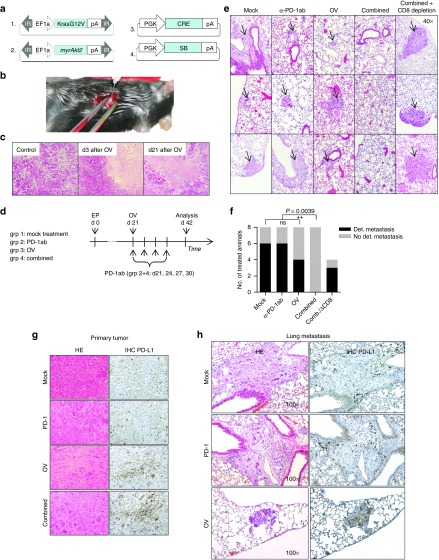
There is evidence that viral oncolysis is synergistic with immune checkpoint inhibition in cancer therapy but the underlying mechanisms are unclear. Here, we investigated whether local viral infection of malignant tumors is capable of overcoming systemic resistance to PD-1-immunotherapy by modulating the spectrum of tumor-directed CD8 T-cells. To focus on neoantigen-specific CD8 T-cell responses, we performed transcriptomic sequencing of PD-1-resistant CMT64 lung adenocarcinoma cells followed by algorithm-based neoepitope prediction. Investigations on neoepitope-specific T-cell responses in tumor-bearing mice demonstrated that PD-1 immunotherapy was insufficient whereas viral oncolysis elicited cytotoxic T-cell responses to a conserved panel of neoepitopes. After combined treatment, we observed that PD-1-blockade did not affect the magnitude of oncolysis-mediated antitumoral immune responses but a broader spectrum of T-cell responses including additional neoepitopes was observed. Oncolysis of the primary tumor significantly abrogated systemic resistance to PD-1-immunotherapy leading to improved elimination of disseminated lung tumors. Our observations were confirmed in a transgenic murine model of liver cancer where viral oncolysis strongly induced PD-L1 expression in primary liver tumors and lung metastasis. Furthermore, we demonstrated that combined treatment completely inhibited dissemination in a CD8 T-cell-dependent manner. Therefore, our results strongly recommend further evaluation of virotherapy and concomitant PD-1 immunotherapy in clinical studies.


Figures

Figure 1
Figure 1
Oncolytic virotherapy elicits specific CD8 T-cell responses against a defined panel of neoepitopes in PD-1-resistant CMT64 tumors. (a) Work flow for prediction and identification of neoepitope-specific CD8 T-cell responses in CMT64 cells. (b) C57BL/6 mice with established s.c. CMT64-tumors were treated twice with αPD-1 antibody (days 0 and 3) or with intratumoral injection of hTertAd. Tumor-free and tumor-bearing mice without treatment served as controls. Splenocytes were harvested 1 week after the first treatment and used for analysis of neoepitope-specific CD8 T-cell responses. For this screen, 44 minimal epitopes from CMT64 sequencing were synthesized as minimal peptides to detect neoepitope-specific responses in IFNγ-ELISpot assays. CD8 T-cell responses observable in all investigated individuals are marked by an asterisk. Responses that were restricted to a subset of mice are marked with “s”. The graph shows representative results from three mice of a total number of eight (n = 8 per group). (c) Ndufs1-V491A-specific CD8 T-cell responses were determined in tumor-free mice treated with a PD-1 blocking antibody and compared to responses in CMT64-tumor-bearing mice (n = 5 per group). Irrelevant peptides served as control. (d) To investigate the ability of neoepitope-specific CD8 T-cell responses to distinguish between tumor-associated neoepitopes and corresponding wild-type epitopes, splenocytes from mice after virotherapy treatment were stimulated with mutated epitope motifs or with corresponding wild-type peptides, respectively. Pooled splenocytes were used for analysis (n = 5 animals). (e) Tabular overview containing a list of detected virotherapy-responsive neoepitopes and additional key information.
Figure 2
Figure 2
Intratumoral inflammation by toll-like receptor (TLR)-ligands is unable to trigger neoepitope-specific CD8 T-cell responses. (a) To investigate whether TLR-ligands can mimic the immunogenicity of viral oncolysis and are capable of triggering tumor-specific CD8 T-cell responses upon injection, mice received intratumoral treatment with the TLR-ligands CpG or poly(I:C), and oncolytic virus as positive control. The CMT64-derived peptide library was used to screen for putative tumor-responses by ELISpot analysis. Representative results of tumor-specific responses from three out of five mice treated with CpG-ODN are shown. (b) Likewise, the graph shows responses from three representative out of five animals treated with poly(I:C). (c) The figure shows ELISpot results from a mouse receiving intratumoral oncolytic virotherapy for the purpose of comparison.
Figure 3
Figure 3
Virotherapy triggers cytotoxic neoepitope-specific CD8 T-cell responses and efficiently eradicates uninfected lung metastases. (a) C57BL/6 mice with subcutaneous CMT64 tumors were treated with or without oncolytic virotherapy. To determine cytotoxic activity of tumor-specific CD8 T-cells, an in vivo cytotoxic assay was performed by adoptive transfer of donor cells pulsed with a peptide pool consisting of all seven tumor-reactive epitopes triggered by oncolytic virotherapy. Cytotoxicity was calculated from ratios of the CFSEhi-target peak pulsed with the tumor-specific peptide pool and the CFSElo-reference peak pulsed with irrelevant peptides. The virus-specific response to dbp served as positive control. Representative histograms are shown in the left panel. The right panel shows the calculated cytotoxicity of indicated groups. (b) Antitumoral efficacy of tumor-specific CD8 T-cells was investigated by clearance of pre-established CMT64 lung colonies. Mice received an i.v. injection of CMT64 cells to establish lung colonies and were treated with intratumoral injections of hTertAd. On day 18 after therapy, mice were sacrificed and lungs were prepared for histology. Untreated mice served as control. To assess the contribution of CD8 T-cells to the clearance of lung colonies, an additional group received virotherapy and CD8 depleting antibodies. The tumor-area of colonies was calculated by computer-based analysis from HE-stained lung sections. The ratio of tumor-area to total lung area is shown in the graph on the right side.
Figure 4
Figure 4
PD-1 inhibition during virus-mediated tumor inflammation broadens the epitope spectrum of neoantigenome-directed CD8 T-cell responses. (a) Using Ndufs1-V491A-specific pentamers for direct T-cell receptor staining, tumor-directed CD8 T-cell responses were monitored in mice after PD-1 checkpoint inhibition, oncolytic virotherapy, and a combination of both treatments. Tumor-free mice and untreated CMT64-tumor bearing mice served as controls. The gating in the representative dotplots (left panel) refers to Ndufs1-V491A-specific CD8 T-cells and the corresponding quantitative analysis is shown on the right panel. (b) Mice bearing s.c. CMT64-tumors received a combination of PD-1 checkpoint blockade (d0 and d3) and oncolytic virotherapy (n = 8). 7 days following virotherapy, mice were screened for neoantigenome-specific CD8 T-cell responses. Epitopes induced by virotherapy are marked by asterisks. Additionally detected T-cell responses, which were neither induced by PD-1 blockade nor by virotherapy alone, are indicated by arrows. Additional epitopes, which were observable in at least 50% of responding individuals are marked by a plus sign. (c) For each treatment group, CD45+ CD11c+ leukocytes were prepared from tumor-draining lymphnodes (TLN) and nontumor-draining lymphnodes (NLN) and PD-L1 expression was quantified by flow cytometry. For those cells derived from corresponding treatment groups, mean fluorescence intensity of PD-L1 is indicated in the graph on the left side. The right panel shows representative histograms. (d) Accordingly, tumor-resident CD45+ CD11c+ leukocytes were prepared from CMT64 tumor-tissue and PD-L1 expression was analyzed.
Figure 5
Figure 5
PD-1 checkpoint inhibition during virus-mediated tumor inflammation results in improved eradication of lung colonies. (a) Therapeutic efficacy of combined treatment with α-PD-1 ab and oncolytic virotherapy was assessed in a subcutaneous model of CMT64 including an increased intrapulmonary burden of metastasis (mice received an intravenous injection of 8 × 105 cells). The experimental timeline and the therapeutic groups are shown. Mice were treated by intratumoral virotherapy and/or α-PD-1 ab as described in the method section. (b) Mice were sacrificed on day 18 after development of lung colonies. Therapeutic efficacy was investigated histologically by measurement of the tumor area as described above. Combination therapy was significantly more efficient than the most effective monotherapy (virotherapy P < 0.0188). (c) Representative lung histologies are shown. (d) Therapeutic efficacy of combined treatment with αPD-1 antibody and oncolytic virotherapy was assessed by survival monitoring. Wild-type mice treated with both therapies demonstrate significantly prolonged survival compared to monotherapies. (e) The graph shows application of the same therapeutic scheme in CD8 knockout mice.
Figure 6
Figure 6
Viral oncolysis and concomitant PD-1 immunotherapy leads to complete elimination of spontaneous lung metastasis in a transgenic model of cholangiocarcinoma. (a) Transposon-based plasmids coding for KrasG12V and myrAkt2 and plasmids expressing SB13-transposase and Cre recombinase were used to establish locally restricted liver tumors by in-situ electroporation. On day 21 after tumor induction, mice developed a single nodule of cholangiocarcinoma suitable for intratumoral injections. (b) The photo illustrates the intratumoral application of virotherapy to the electroporation-induced liver tumor in situ. (c) Tumors were treated with intratumoral injection of hTertAd. On day 3 and day 21 after virotherapy, intratumoral lysis was investigated on HE-stained sections at 40-fold magnification. (d) Mice bearing intrahepatic tumors received a systemic administration of PD-1-blocking antibodies, intratumoral virotherapy, or a combination of both (n = 8 animals per group). For virotherapeutic application, mice were laparotomized and received intatumoral injections of virotherapy, or physiologic salt solution as control, respectively. Twenty-one days after treatment, lungs were screened for metastases. (e) Three representative lung histologies from each group are shown. (f) The graph displays the distribution of mice with detectable metastasis and metastasis-free mice for each treatment group according to the scheme shown in Figure 6d. An additional group treated with combined therapy received CD8-depleting antibodies (n = 4 animals). PD-L1 expression in primary tumors (g) and metastases (h) after different therapies as indicated was investigated by HE stainings and immunohistochemical staining (100-fold magnification).

References

    1. Topalian, SL, Hodi, FS, Brahmer, JR, Gettinger, SN, Smith, DC, McDermott, DF et al. (2012). Safety, activity, and immune correlates of anti-PD-1 antibody in cancer. N Engl J Med 366: 2443–2454. - PMC - PubMed
    1. Hodi, FS, O'Day, SJ, McDermott, DF, Weber, RW, Sosman, JA, Haanen, JB et al. (2010). Improved survival with ipilimumab in patients with metastatic melanoma. N Engl J Med 363: 711–723. - PMC - PubMed
    1. Brahmer, JR, Tykodi, SS, Chow, LQ, Hwu, WJ, Topalian, SL, Hwu, P et al. (2012). Safety and activity of anti-PD-L1 antibody in patients with advanced cancer. N Engl J Med 366: 2455–2465. - PMC - PubMed
    1. Lynch, TJ, Bondarenko, I, Luft, A, Serwatowski, P, Barlesi, F, Chacko, R et al. (2012). Ipilimumab in combination with paclitaxel and carboplatin as first-line treatment in stage IIIB/IV non-small-cell lung cancer: results from a randomized, double-blind, multicenter phase II study. J Clin Oncol 30: 2046–2054. - PubMed
    1. Powles, T, Eder, JP, Fine, GD, Braiteh, FS, Loriot, Y, Cruz, C et al. (2014). MPDL3280A (anti-PD-L1) treatment leads to clinical activity in metastatic bladder cancer. Nature 515: 558–562. - PubMed

Publication types

MeSH terms