. 2015 Jun 26:6:7481.
doi: 10.1038/ncomms8481.

Glycan complexity dictates microbial resource allocation in the large intestine

Affiliations

Glycan complexity dictates microbial resource allocation in the large intestine

Artur Rogowski et al. Nat Commun. .

Erratum in

Abstract

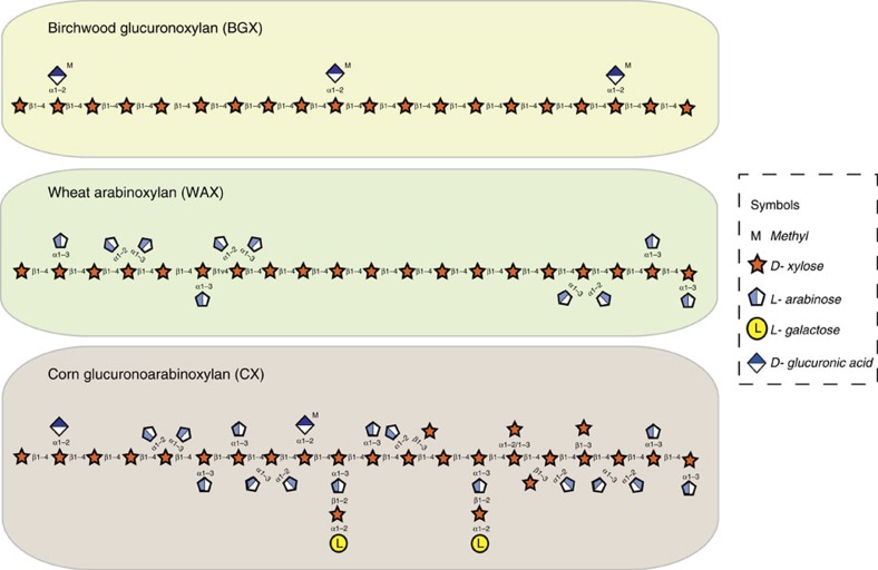
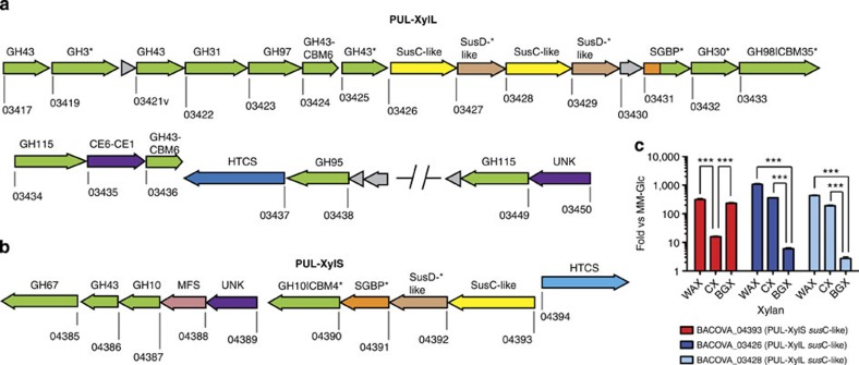
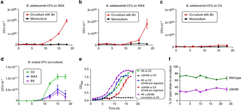
The structure of the human gut microbiota is controlled primarily through the degradation of complex dietary carbohydrates, but the extent to which carbohydrate breakdown products are shared between members of the microbiota is unclear. We show here, using xylan as a model, that sharing the breakdown products of complex carbohydrates by key members of the microbiota, such as Bacteroides ovatus, is dependent on the complexity of the target glycan. Characterization of the extensive xylan degrading apparatus expressed by B. ovatus reveals that the breakdown of the polysaccharide by the human gut microbiota is significantly more complex than previous models suggested, which were based on the deconstruction of xylans containing limited monosaccharide side chains. Our report presents a highly complex and dynamic xylan degrading apparatus that is fine-tuned to recognize the different forms of the polysaccharide presented to the human gut microbiota.


Figures

Figure 1
Figure 1. Schematic of the structures of the main classes of xylan.
The monosaccharides and linkages in the main classes of xylan are shown and are represented in their Consortium for Functional Glycomics format. The xylans used in this study were from birchwood (birch glucuronoxylan; BGX), wheat flour (wheat arabinoxylan; WAX) and corn bran (corn glucuronoarabinoxylan; CX).
Figure 2
Figure 2. Schematic of the B. ovatus xylan PULs and differential expression during growth on different xylans.
(a,b) schematic of PUL-XylL and PUL-XylS, respectively. Each gene is drawn to scale as a rectangle with its orientation indicated by the arrow head. The numbers below each gene is its locus tag (bacova_XXXXX). Genes encoding known or predicted functionalities are colour-coded and, where appropriate, are also annotated according to their CAZy family number: glycoside hydrolase (GH; green), carbohydrate esterase (CE; purple), carbohydrate-binding module (CBM). Surface located proteins are marked with an asterisk. SGBP=surface glycan binding proteins are coloured orange, or, if also in a CAZy family (BACOVA_03431; inactive GH10), are coloured half orange, half green. UNK=unknown (purple), but distant similarity to CE6 carbohydrate esterases. HTCS=hybrid two component system (light or dark blue). MFS=transporter of the major facilitator superfamily (pink). Grey=unknown function (note, there is a structure of BACOVA_03430, PDB accession code 3N91). SusC-like (yellow) and SusD-like (light tan) proteins are a defining feature of PULs and are responsible for import of complex glycans across the outer membrane. SusC-like proteins are TonB-dependent transporters, while SusD-like proteins are surface lipoproteins that likely function to deliver the target glycan to their partner SusC. (c) Cells were grown on minimal media with polysaccharide as the sole carbon source, and levels of different susC transcripts (locus tags shown; used as a proxy for expression of the whole PUL) from each PUL were quantified by qRT-PCR. The y-axis shows the Log fold-change relative to a minimal media-glucose reference; x-axis labels indicate the xylans used. WAX= wheat arabinoxylan, CX=corn xylan, BGX=birchwood glucuronoxylan. CX is a highly complex xylan compared to WAX and BGX. All data were analysed by one-way ANOVA followed by Tukey's multiple comparison test (***=P≤0.001).
Figure 3
Figure 3. Cellular location of Xylan PUL enzymes containing a predicted N-terminal lipidation site or uncleaved transmembrane anchor.
(a) Fluorescent microscopic images of B. ovatus cells cultured on WAX and incubated with polyclonal antibodies raised against recombinant BACOVA_03419 (GH3), BACOVA_03421 and BACOVA_03425 (GH43s), BACOVA_03431 (SGBP), BACOVA_03432 (GH30), BACOVA_03433 (GH98) and BACOVA_04390 (GH10). (b) Western blots of the B. ovatus cells shown in panel a, either untreated with Proteinase K (-) or incubated with 2 mg ml−1 Proteinase K for 16 h (+). The blots were probed with antibodies against the B. ovatus proteins indicated. The full blots are shown in Supplementary Fig. 8.
Figure 4
Figure 4. The activity of the GH98 enzyme BACOVA_03433.
(a) Different xylans at 1% (w/v) were incubated with 1 μM of the GH98 enzyme in PBS for 16 h at 37 °C. The reaction products were subjected to thin layer chromatography and compared with xylooligosaccharide standards with a d.p. ranging from 1 to 5 (X1-X5). (b) HPAEC-PAD trace showing the high molecular weight oligosaccharide products of CX digestion by the GH98 endo-xylanase, separated using a sodium acetate gradient of 0-500 mM. The reaction conditions used are the same as in panel a Control reactions of substrate and enzyme only are shown. (c) CX was incubated with 5 μM of the GH98 enzyme for 24 h in 50 mM sodium phosphate buffer, pH 7.0, at 37 °C. The reaction products, and CX that was not incubated with the GH98 enzyme, were then treated with sodium borohydride, to convert the reducing end monosaccharide unit into its alditol, followed by acid hydrolysis to convert the oligosaccharides (or in the case of control CX alone) into their monosaccharide constituents. The hydrolysed products were subjected to HPAEC and compared to the migration of standard monosaccharides as well as the alditol of xylose (xylitol) and galactose (galactitol). (d) The GH98 enzyme was incubated with CX (lane CX vs GH98), or CX that had been pre-treated with all the side chain cleaving enzymes encoded by PUL-XylL (lane CX vs MIX), or CX that had been pre-treated with all the side chain cleaving enzymes encoded by PUL-XylL, except the GH43 enzyme BACOVA_03417 that removes O3 linked arabinose from double substituted xylose (lane CX vs MIX no double abfase).
Figure 5
Figure 5. Activity and structure of BACOVA_03438 GH95 α-L-galactosidase and comparison to the Bifidobacterium bifidum GH95 α-L-fucosidase.
(a,b) TLC and HPAEC profiles, respectively, of the reaction product (L-galactose) generated by incubating CX at 1 mg ml−1 with 1 μM BACOVA_03438 for 16 h in 20 mM sodium phosphate buffer, pH 7.0, at 37 °C. In (a) the lanes are as follows: (−) CX no enzyme; (+) CX incubated with BACOVA_03438. The asterisk marks the position of the single reaction product released from CX by the GH95. (c) The product of the GH95 is resistant to oxidation by D-galactose dehydrogenase (GDH). (d) Schematic of BACOVA_03438, colour-ramped from blue (N-terminus) to red (C-terminus), with L-Gal bound in the active site (carbohydrate carbons shown as green sticks). (e) Solvent exposed surface representation of B. ovatus GH95. The active site pocket that houses β-L-Gal product is boxed. (f) Side chains of the active site residues of BACOVA_03438 (magenta carbons) and the GH95 α-L-fucosidase (blue carbons; PDB: 2EAE) from Bifidobacterium bifidum, bound to L-Gal (green carbons) and L-Fuc (orange carbons), respectively. Predicted hydrogen bonds between the amino-acid side chains and carbohydrates are shown as yellow dotted lines, except for the H-bond between the Oγ of BACOVA_03438 Thr370 and O6 of L-Gal, which is shown as a red dotted line. (g) Overlay of the active site residues displayed in panel (f) with the side-chains of the Thr/His polymorphism that may play a role in specificity for L-Gal over L-Fuc labelled. A stereo image of a portion of the electron density map is shown in Supplementary Fig. 9.
Figure 6
Figure 6. Growth profile of wild type and mutants of B. ovatus on xylans.
WT and PUL deletion mutants were cultured in minimal media containing different xylans at 0.5% (w/v) as the sole carbon source. Birchwood glucuronoxylan (BGX; (a)) and wheat arabinoxylan (WAX; (b)) are relatively simple, sparsely decorated structures, while corn (c), sorghum (d) and rice (e) xylans are more complex heavily decorated glucuronoarabinoxylans (GAXs). The high starting OD for BGX is due to the background turbidity of the polysaccharide. A total of 6 replicate cultures were monitored at 20 min intervals for each substrate and used to generate the average growth curves shown.
Figure 7
Figure 7. Models for the degradation of different forms of xylan by B. ovatus.
In the upper part of each panel the monosaccharides and linkages in the main classes of xylan are shown and are represented in their Consortium for Functional Glycomics format. The lower parts of each panel show the model for degradation of complex GAXs like CX (a) AXs like WAX (b) and GXs like BGX (c) The red asterisk next to BACOVA_03425 GH43 indicates that the single specific arabinofuranosidase acts after BACOVA_03417 (α1,3 double-specific arabinofuranosidase) to remove the remaining α1,2 linked L-arabinose. The black arrows indicate examples of the linkages cleaved by the enzymes, shown in green ovals and identified by their glycoside hydrolase family and locus tag. In panel a, the dotted arrows from the GH98 enzyme identify possibly linkages hydrolysed by the xylanase. Although not shown on the CX model, the PUL-XylS apparatus will be present, albeit at low levels (see Fig. 2c). A schematic of the crystal structures, colour-ramped from blue (N-terminus) to red (C-terminus), of two of the xylan degrading enzymes, determined in this study (BACOVA_03438 GH95; PDB accession code 4UFC) and in a recent report (BACOVA_03449 GH115; PDB accession code 4C91) are shown. The red arrow heads indicate glycan transport between cellular locations, while the yellow arrow heads show the ligand (or potential ligand in case of the question mark) that binds and activates the hybrid two component system (HTCS) encoded by PUL-XylS (BACOVA_04394, light blue HTCS; activating ligand xylotetraose4) or PUL-XylL (BACOVA_03437, dark blue HTCS; activating ligand likely an arabino-xylooligosaccharide). MFS is a member of the Major Facilitator Superfamily of transport proteins, and SGBP signifies the two surface xylan binding proteins encoded by the xylan PULs, BACOVA_03431 (inactive GH10, PUL-XylL) and BACOVA_04391 (PUL-XylS). A schematic of the crystal structure of BACOVA_04391 (PDB accession code 3ORJ), colour-ramped from blue (N-terminus) to red (C-terminus), is shown. Note only one of the two SusC/D pairs from PUL-XylL is shown for clarity.
Figure 8
Figure 8. Conservation of the B. ovatus ATCC 8483 xylan PULs in other Bacteroides species.
CAZymes (green, CBM modules were omitted for clarity), susC-like (yellow), susD-like (orange) and the HTCS sensor-regulator (blue) genes are colour-coded. Genes encoding proteins of unknown function or non-CAZymes (for example, surface glycan binding protein and MFS carbohydrate importer in PUL-XylS) are shown in grey. (a) PUL-XylL. Vertical dotted lines indicate split gene models, while missing gene models are shown in red. Microsyntenic segments are depicted by grey connectors, and include adjacent conserved non-PUL members (shown with dotted lines). Eighteen genes are conserved across all strains, and 17 of these form three highly conserved blocks indicated on top. The major difference between the two groups of the GH98 for GH5_21 swap is indicated. Inset shows a TLC of the activity of the recombinant B. xylanisolvens XBA1 GH5_21 enzyme (BXY_29320; boxed in red) against different xylans (1 μM final enzyme vs 0.5% xylan in 20 mM sodium phosphate buffer, pH 7.0, 16 h at 37 °C). The B. xylanisolvens GH5 enzyme displays endo-xylanase activity against CX, producing a smear of large oligosaccharide products similar to the product profile observed for the B. ovatus GH98 enzyme, BACOVA_03438. The GH5 xylanase also hydrolyses WAX, unlike the GH98 enzyme, but displays no activity against BGX. X1-X5 indicates xylooligosaccharide standards. (b) PUL-XylS. The lower part of the panel shows the strains that contain only the second half of PUL-XylS that encodes the intracellular (periplasmic and cytoplasmic) enzymes only.
Figure 9
Figure 9. The ability of other members of the microbiota to use PBPs released by B. ovatus during growth on xylan is determined by the complexity of the polysaccharide.
Bacteroides ovatus (Bo) and Bifidobacterium adolescentis were co-cultured on BGX (a) WAX (b) and CX (c) and the number of CFUs of each determined at different points on the growth curve. (d) Shows the CFU ml-1 of B. ovatus alone at different phases of growth. Note, when the Bifidobacterium alone was grown on digested xylans (see Supplementary Fig. 8), the CFU ml−1 at late exponential phase (OD600 ∼1.2) was ∼8.0 × 10⁸, indicating that ∼25% of the total xylan is used by B. adolescentis. (e) Growth of B. ovatus wild type and the ΔGH98 mutant (BACOVA_03433) on CX (0.5% w/v). Pre-digested indicates the CX was digested to completion with the GH98 xylanase prior to addition to the media. (f) Tagged strains of wild-type B. ovatus and the ΔGH98 mutant were co-cultured on CX (see panel e for growth curve). Samples were taken at different time points and qPCR with primers unique to each strain was used to determine the ratio of each in the culture. Each data point is the average and s.d. of triplicate growths.

References

    1. Arumugam M. et al. Enterotypes of the human gut microbiome. Nature 473, 174–180 (2011). - PMC - PubMed
    1. Backhed F., Ley R. E., Sonnenburg J. L., Peterson D. A. & Gordon J. I. Host-bacterial mutualism in the human intestine. Science 307, 1915–1920 (2005). - PubMed
    1. Martens E. C., Chiang H. C. & Gordon J. I. Mucosal glycan foraging enhances fitness and transmission of a saccharolytic human gut bacterial symbiont. Cell Host Microbe 4, 447–457 (2008). - PMC - PubMed
    1. Martens E. C. et al. Recognition and degradation of plant cell wall polysaccharides by two human gut symbionts. PLoS Biol. 9, e1001221 (2011). - PMC - PubMed
    1. Flint H. J., Scott K. P., Duncan S. H., Louis P. & Forano E. Microbial degradation of complex carbohydrates in the gut. Gut Microbes 3, 289–306 (2012). - PMC - PubMed

Publication types

LinkOut - more resources