. 2015 Jun 26:6:7494.
doi: 10.1038/ncomms8494.

Co-translational capturing of nascent ribosomal proteins by their dedicated chaperones

Affiliations

Co-translational capturing of nascent ribosomal proteins by their dedicated chaperones

Patrick Pausch et al. Nat Commun. .

Abstract

Exponentially growing yeast cells produce every minute >160,000 ribosomal proteins. Owing to their difficult physicochemical properties, the synthesis of assembly-competent ribosomal proteins represents a major challenge. Recent evidence highlights that dedicated chaperone proteins recognize the N-terminal regions of ribosomal proteins and promote their soluble expression and delivery to the assembly site. Here we explore the intuitive possibility that ribosomal proteins are captured by dedicated chaperones in a co-translational manner. Affinity purification of four chaperones (Rrb1, Syo1, Sqt1 and Yar1) selectively enriched the mRNAs encoding their specific ribosomal protein clients (Rpl3, Rpl5, Rpl10 and Rps3). X-ray crystallography reveals how the N-terminal, rRNA-binding residues of Rpl10 are shielded by Sqt1's WD-repeat β-propeller, providing mechanistic insight into the incorporation of Rpl10 into pre-60S subunits. Co-translational capturing of nascent ribosomal proteins by dedicated chaperones constitutes an elegant mechanism to prevent unspecific interactions and aggregation of ribosomal proteins on their road to incorporation.


Figures

Figure 1
Figure 1. Sqt1 and Rrb1 recognize the N-terminal residues of Rpl10 and Rpl3, respectively.
(a) Sqt1 and Rrb1 are exclusively associated with Rpl10 and Rpl3. TAP of C-terminally TAP-tagged Sqt1 (Sqt1-TAP, lane 1) and N-terminally TAP-tagged Rrb1 (NTAP-Rrb1, lane 2) from yeast cell lysates. Final EGTA eluates were analysed by SDS–polyacrylamide gel electrophoresis (PAGE) and Coomassie staining. (b) Y2H interaction between Rpl10 and Sqt1. Note that the Sqt1.53C protein lacks amino acids 1–52 and thus essentially contains the WD-repeat β-propeller domain of Sqt1. Rpl10.12C corresponds to an Rpl10 variant starting with amino acid 12. (c) Y2H interaction between Rpl3 and Rrb1. Note that the Rrb1.60C protein lacks amino acids 2–59 and thus contains the WD-repeat β-propeller domain, including a predicted N-terminal α-helix, of Rrb1 (Supplementary Fig. 2d). Rpl3.8C corresponds to an Rpl3 variant starting with amino acid eight. (d) In vitro binding assay between Rpl10 and Sqt1. The indicated C-terminally (His)6-tagged Rpl10 and C-terminally Flag-tagged Sqt1 variants were co-expressed in E. coli and purified via Ni-affinity purification. Proteins were revealed by SDS–PAGE and Coomassie staining (top) or by western blot analysis using anti-Flag (Sqt1-Flag variants) and anti-His (Rpl10-(His)6 variants) antibodies (bottom). T, total extract (lane 1); P, pellet fraction (insoluble proteins, lane 2); S, soluble extract (lane 3); E, imidazole eluate (lane 4); M, molecular weight standard. The bands highlighted by blue arrowheads correspond to the different Rpl10 variants used as baits for the purifications. Black arrowheads indicate the position of Sqt1-Flag and Sqt1.53C-Flag. Note that the third panel can be considered as a reference for the background binding of Sqt1-Flag to the Ni-NTA agarose resin.
Figure 2
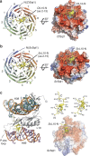
Figure 2. Crystal structures of the eight-bladed WD-repeat β-propeller domain of Sqt1 with bound L10-N from S. cerevisiae and C. thermophilum.
(a) Crystal structure of ctSqt1.52C (residues 52–533) with bound ctL10-N (residues 2–13). Cartoon representation showing ctSqt1.52C in rainbow colours from N- to C terminus and ctL10-N in yellow with side chains (left panel). The eight-bladed WD-repeat β-propeller is shown in its top view. Assignment of the top and bottom surface as well as numbering of the propeller blades (1–8) and labelling of the β-strands within each blade (a–d) is according to the conventional definition for WD-repeat β-propellers. N- and C termini are indicated. Electrostatic properties of the top surface of ctSqt1.52C with bound L10-N in yellow (right panel). (b) Crystal structure of ScSqt1.53C (residues 53–431) with bound ScL10-N (residues 2–15). Cartoon representation (left panel) and electrostatic properties (right panel) of ScSqt1.53C with bound ScL10-N in yellow. Labels and colouring is as in a. (c) Comparison of the interaction modes of ScL10-N with helix H89 of the 25S rRNA and with ScSqt1, respectively. Cartoon representation of Rpl10 bound to H38 and H89 of the 25S rRNA as observed in the mature 60S subunit (PDB 3U5I and 3U5H for Rpl10 and 25S rRNA, respectively) (left panel) and bound to ScSqt1.53C (right panel). The N-terminal residues of Rpl10 (amino acids 2–15) are shown in yellow with side chains, the remainder of Rpl10 in grey, and bases of H38 in turquoise and of H89 in purple (phosphate backbones of H38 and H89 are shown in orange). Sqt1 is shown in its surface representation with electrostatic properties. The upper right part shows a comparison of the L10-N peptide in the ribosome-bound (left) and Sqt1-bound (right) state.
Figure 3
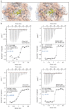
Figure 3. A thermophilic adaptation might sense the processing status of Rpl10's N-terminal methionine.
(a) Close-up of the interaction between the L10-N residues and the WD-repeat β-propeller domain of CtSqt1 (left panel) and of ScSqt1 (right panel). Sqt1 is shown in its cartoon representation with superimposed electrostatic surface properties. The Sqt1 residues involved in the interaction are shown as sticks. L10-N residues (yellow) are shown in a mixed cartoon/stick representation. The relevant Sqt1 and L10-N residues are labelled in blue and black, respectively (for example, A2 for Ala2). N- and C termini of L10-N are indicated (N′ and C′). (b) Analysis of the L10-N interaction with the WD-repeat β-propeller domain of CtSqt1 and ScSqt1 by ITC. Shown are ITC measurements of CtSqt1.52C/ScSqt1.53C with CtL10-N/ScL10-N peptides either lacking (amino acids 2–20) or including Met1 (amino acids 1–20), as indicated in each panel. The upper part of each panel shows the raw injection heats (μcal s−1). The lower part of each panel displays the corresponding specific binding isotherms (Kcal mol−1 of injectant) plotted against the molar ratio. The measured interaction parameters are listed within the profiles and the approximate Kd is shown in blue.
Figure 4
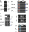
Figure 4. Ionic interactions are critical determinants of L10-N binding by Sqt1.
(a) Representation of the mode of L10-N recognition by ScSqt1. The backbone and side chains of ScL10-N (residues 2–13) are shown as an elongated peptide. L10-N residues are labelled in grey (e.g.,: A2 for Ala2). The Sqt1 residues that form interactions, either via their side chains or main-chain carbonyls, with the L10-N peptide are indicated. Dotted lines indicate ionic interactions or hydrogen bonds and grey, curved lines hydrophobic interactions. The interaction representation was created with Accelrys Draw 4.1. (b) Y2H interaction between Sqt1 and Rpl10 variants harbouring mutations within the N-terminal residues. The residues mutated in Rpl10 (for example, R3E for Arg3 to glutamate), as well as the Sqt1 residues they are contacting (blue arrowheads, Sqt1*), are indicated. (c) Y2H interaction between Rpl10 and mutant Sqt1 variants. The residues mutated in Sqt1 (for example, E110K for Glu110 to lysine), as well as the L10-N residues they are contacting (blue arrowheads, Rpl10*), are indicated. Single-letter abbreviations for the amino acid residues are as follows: A, Ala; D, Asp; E, Glu; I, Ile; K, Lys; R, Arg; T, Thr; and Y, Tyr.
Figure 5
Figure 5. Overexpression of Rpl10 bypasses the requirement for the essential Sqt1.
(a) In vivo phenotypes of cells expressing Sqt1 variants that affect the interaction with Rpl10. Growth phenotypes of viable sqt1 mutants on YPD plates that were incubated for 2 days at 30 °C (left panel). The lethality of sqt1 alleles that abolish the interaction with Rpl10 was scored on plates containing 5-fluoroorotic Acid (5-FOA), which were incubated for 3 days at 30 °C (right panel). The residues mutated in Sqt1, as well as the L10-N residues they are contacting (blue arrowheads, Rpl10*), are indicated. (b) Overexpression of Rpl10 suppresses the slow-growth phenotype of cells expressing Sqt1 variants that affect the interaction with Rpl10 and even rescues the absence of Sqt1. Cells harbouring the SQT1 wild-type allele or the indicated sqt1 alleles were grown in the absence (vector) or presence of a multicopy plasmid expressing Rpl10 (RPL10) on SC-Leu-Trp plates that were incubated for 3 days at 30 °C. (c) In vivo phenotypes of cells expressing Rpl10 variants harbouring mutations within the N-terminal residues. The growth phenotypes of the indicated rpl10 alleles were scored on YPD plates (viable mutants; upper panel), which were incubated as indicated, and on 5-FOA-containing plates (lethal mutants; lower panel), which were incubated for 3 days at 30 °C.
Figure 6
Figure 6. The essential function of Sqt1 consists in Rpl10 binding.
(a) Allele-specific synthetic lethality between interaction surface mutants of sqt1 and rpl10. The growth phenotypes of cells harbouring the SQT1 wild-type allele or the indicated sqt1 alleles in combination with the RPL10 wild-type allele or the rpl10.R3E and rpl10.R4A allele were scored on 5-FOA-containing plates, which were incubated for 4 days at 30 °C. The mutated Sqt1 residues, as well as the L10-N residues they are contacting (blue arrowheads, Rpl10*), are indicated. (b) Allele-specific abrogation of the interaction between the above interaction surface mutants of sqt1 and rpl10. Y2H interactions were assessed for combinations between Sqt1 or the indicated Sqt1 variants and Rpl10 or the Rpl10.R3E and Rpl10.R4A mutant variant.
Figure 7
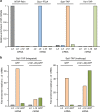
Figure 7. Chaperones are recruited to nascent ribosomal proteins.
(a) The chaperone proteins were affinity purified (IgG-Sepharose pull-down) from extracts of cycloheximide-treated cells and the associated RNA was isolated from the TEV eluates. Each of the four chaperone purifications (NTAP-Rrb1, Syo1-FTpA, Sqt1-TAP, and Yar1-TAP) was assessed for their content of the four ribosomal protein (RP) mRNAs (RPL3, RPL5, RPL10 and RPS3) by real-time qRT–PCR. The data from one representative experiment are expressed as the relative enrichment of the specifically co-purified RP mRNA in each of the four chaperone purifications (see Methods section for details). For each cDNA, real-time qPCRs were performed in triplicates. A highly reproducible data set was obtained in an independent series of chaperone purifications. (b) The N-terminal residues of Rpl10 are sufficient to target Sqt1 to the nascent Rpl10(1–20)-yEGFP fusion protein. Sqt1-TAP, either expressed from the genomic locus (left panel) or from a multicopy plasmid (right panel) was affinity purified (IgG-Sepharose pull-down) from extracts of cells where expression of either the yEGFP (GFP) control protein or the Rpl10(1–20)-yEGFP [L10(1–20)-GFP] fusion protein has been induced from the CUP1 promoter for 10 min with 500 μM copper sulfate. The Sqt1-TAP purifications were assessed for their content of the RPL3, RPL10 and yEGFP (GFP) mRNAs by real-time qRT–PCR. The data from one representative experiment are expressed as the fold enrichment relative to the RPL3 mRNA. For each cDNA, real-time qPCRs were performed in triplicates. Note that the bar graphs of the left and right panel of this figure are at a different scale.
Figure 8
Figure 8. Model highlighting the co-translational capturing of selected ribosomal proteins by their dedicated chaperones.
Simplified model for the stable incorporation of Rpl10 into cytoplasmic pre-60S subunits (upper left part). The chaperone Sqt1 recognizes the N-terminal residues of Rpl10 as these emerge from translating ribosomes and the Sqt1–Rpl10 complex is released into the cytoplasm upon translation termination. We propose that initial docking of Sqt1-bound Rpl10 onto Lsg1-defined pre-60S subunits involves Rpl10 surfaces that are not shielded by Sqt1 and likely occurs at pre-60S sites that are not masked by Nmd3 (see Discussion section). Subsequently, the activity of the GTPase Lsg1 entails structural rearrangements that promote the release of Nmd3 and the stable incorporation of Rpl10, thus leading to the generation of mature 60S subunits that can engage in translation initiation. General model for the co-translational capturing of ribosomal proteins by their specific chaperone partners (lower left part). This study has revealed that the chaperones Rrb1, Syo1 and Yar1 are also recruited to their distinct ribosomal protein clients (Rpl3, Rpl5 and Rps3), as these are synthesized from their mRNAs by the ribosome. While the Rrb1-Rpl3 and Syo1-Rpl5-Rpl11 complexes are imported into the nucleus where these ribosomal proteins assemble into pre-60S subunits, it is not yet clear whether Yar1 travels together with Rps3 into the nucleus or promotes pre-40S assembly of Rps3 in the cytoplasm. After extensive nuclear maturation (right part), pre-60S particles gain export competence upon recruitment of Nmd3, which is recognized by the exportin Crm1, and travel across the nuclear pore complex to the cytoplasm.

Similar articles

Cited by

References

    1. Ben-Shem A. et al. The structure of the eukaryotic ribosome at 3.0Å resolution. Science 334, 1524–1529 (2011). - PubMed
    1. Klinge S., Voigts-Hoffmann F., Leibundgut M., Arpagaus S. & Ban N. Crystal structure of the eukaryotic 60S ribosomal subunit in complex with initiation factor 6. Science 334, 941–948 (2011). - PubMed
    1. Melnikov S. et al. One core, two shells: bacterial and eukaryotic ribosomes. Nat. Struct. Mol. Biol. 19, 560–567 (2012). - PubMed
    1. Rabl J., Leibundgut M., Ataide S. F., Haag A. & Ban N. Crystal structure of the eukaryotic 40S ribosomal subunit in complex with initiation factor 1. Science 331, 730–736 (2011). - PubMed
    1. Warner J. R. The economics of ribosome biosynthesis in yeast. Trends Biochem. Sci. 24, 437–440 (1999). - PubMed

Publication types

MeSH terms