. 2015 Aug;138(Pt 8):2423-37.
doi: 10.1093/brain/awv154. Epub 2015 Jun 25.

The Wernicke conundrum and the anatomy of language comprehension in primary progressive aphasia

Affiliations

The Wernicke conundrum and the anatomy of language comprehension in primary progressive aphasia

M-Marsel Mesulam et al. Brain. 2015 Aug.

Abstract

Wernicke's aphasia is characterized by severe word and sentence comprehension impairments. The location of the underlying lesion site, known as Wernicke's area, remains controversial. Questions related to this controversy were addressed in 72 patients with primary progressive aphasia who collectively displayed a wide spectrum of cortical atrophy sites and language impairment patterns. Clinico-anatomical correlations were explored at the individual and group levels. These analyses showed that neuronal loss in temporoparietal areas, traditionally included within Wernicke's area, leave single word comprehension intact and cause inconsistent impairments of sentence comprehension. The most severe sentence comprehension impairments were associated with a heterogeneous set of cortical atrophy sites variably encompassing temporoparietal components of Wernicke's area, Broca's area, and dorsal premotor cortex. Severe comprehension impairments for single words, on the other hand, were invariably associated with peak atrophy sites in the left temporal pole and adjacent anterior temporal cortex, a pattern of atrophy that left sentence comprehension intact. These results show that the neural substrates of word and sentence comprehension are dissociable and that a circumscribed cortical area equally critical for word and sentence comprehension is unlikely to exist anywhere in the cerebral cortex. Reports of combined word and sentence comprehension impairments in Wernicke's aphasia come almost exclusively from patients with cerebrovascular accidents where brain damage extends into subcortical white matter. The syndrome of Wernicke's aphasia is thus likely to reflect damage not only to the cerebral cortex but also to underlying axonal pathways, leading to strategic cortico-cortical disconnections within the language network. The results of this investigation further reinforce the conclusion that the left anterior temporal lobe, a region ignored by classic aphasiology, needs to be inserted into the language network with a critical role in the multisynaptic hierarchy underlying word comprehension and object naming.

Keywords: aphasia; dementia; grammar; language; semantics.


Figures

None
The location of Wernicke’s area remains controversial. Using structural MRI in 72 patients with primary progressive aphasia, Mesulam et al. show that word and sentence comprehension are dissociable, and that a circumscribed cortical area equally critical for word and sentence comprehension is unlikely to exist anywhere in the cerebral cortex.
Figure 1
Figure 1
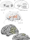
Evolution of Wernicke’s area. (A) From Dejerine’s textbook (Dejerine and Dejerine-Klumpke, 1895). In this figure ‘A’ designates Wernicke’s centre for auditory images of words, ‘B’ designates Broca’s centre for motor images of articulation, and ‘Pc’ designates the centre for visual images of words. (B) From Penfield and Roberts (1959). (C) From Geschwind (1972). (D) Lateral and ventral views of the left hemisphere showing a composite rendition of Wernicke’s area (W, in green). ag = angular gyrus; fg = fusiform gyrus; ifg = inferior frontal gyrus; itg = inferior temporal gyrus; mtg = middle temporal gyrus; phg = parahippocampal gyrus; smg = supramarginal gyrus; stg = superior temporal gyrus; tp = temporal pole of the ATL.
Figure 2
Figure 2
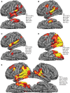
Atrophy maps in Wernicke’s area in Patients P1–7. (A–D) Individual atrophy maps of the left hemisphere and performance in language tasks for four patients with atrophy sites that illustrate the anatomical variability but also the common features of peak atrophy sites within the ‘W’ territory of Fig. 1D but without involving the inferior frontal gyrus or tip of the anterior temporal lobe. (E) Atrophy map in both hemispheres for the whole group. ag = angular gyrus; AQ = Aphasia Quotient of the Western Aphasia Battery; BNT = Boston Naming Test; dpm = dorsal premotor cortex; fg = fusiform gyrus; ifg = inferior frontal gyrus; itg = inferior temporal gyrus; mtg = middle temporal gyrus; phg = parahippocampal gyrus; REP = repetition test; SC = sentence comprehension test; smg = supramarginal gyrus; stg = superior temporal gyrus; tp = temporal pole of the ATL.
Figure 3
Figure 3
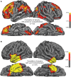
Correlations of cortical thinning (atrophy) with language comprehension scores. FDR threshold was set at 0.05. The heat map numbers indicate −log10 P-values of significance. (A) Sites where atrophy correlates with sentence comprehension impairment. (B) Sites where atrophy correlates with word comprehension impairment when non-verbal object recognition scores are considered nuisance variables. ag = angular gyrus; dpm = dorsal premotor cortex; fg = fusiform gyrus; ifg = inferior frontal gyrus; itg = inferior temporal gyrus; mtg = middle temporal gyrus; phg = parahippocampal gyrus; smg = supramarginal gyrus; stg = superior temporal gyrus; tp = temporal pole of the ATL.
Figure 4
Figure 4
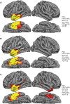
Word comprehension and anterior temporal lobe atrophy. (A–C) Individual atrophy maps and language performance scores in three members of Group P16–25 (the subjects with the 10 lowest word comprehension scores). The three examples display the consistency of the atrophy patterns seen in the group as a whole. ag = angular gyrus; AQ = Aphasia Quotient of the Western Aphasia Battery; BNT = Boston Naming Test; fg = fusiform gyrus; ifg = inferior frontal gyrus; itg = inferior temporal gyrus; mtg = middle temporal gyrus; phg = parahippocampal gyrus; PPTp = picture form of the Pyramids and Palm Trees Test; SC = sentence comprehension test; smg = supramarginal gyrus; stg = superior temporal gyrus; tp = temporal pole of the ATL.
Figure 5
Figure 5
Progression of atrophy in the anterior temporal lobe. (A) Patient P26 has temporopolar atrophy but nearly intact single word comprehension. Severe naming impairment (BNT performance of 40%) stands out as the principal finding. (B) Four years later the atrophy extends further into the anterior temporal lobe and is associated with severe single word comprehension impairment. ag = angular gyrus; AQ = Aphasia Quotient of the Western Aphasia Battery; BNT = Boston Naming Test; fg = fusiform gyrus; ifg = inferior frontal gyrus; itg = inferior temporal gyrus; mtg = middle temporal gyrus; phg = parahippocampal gyrus; PPTp = picture form of the Pyramids and Palm Trees Test; SC = sentence comprehension test; smg = supramarginal gyrus; stg = superior temporal gyrus; tp = temporal pole of the ATL.

Similar articles

Cited by

References

    1. Amici S, Brambati SM, Wilkins DP, Ogar J, Dronkers NF, Miller B, Gorno-Tempini ML. Anatomical correlates of sentence comprehension and verbal working memory in neurodegenerative disease. J Neurosci 2007; 27: 6282–90. - PMC - PubMed
    1. Benson F, Geschwind N. Aphasia and related disorders: a clinical approach. In: Mesulam M-M, editor. Principles of behavioral neurology. Philadelphia, PA: F. A. Davis; 1985. pp. 193–238.
    1. Bogen JE, Bogen GM. Wernicke's region-where is it? Ann N Y Acad Sci 1976; 280: 834–43. - PubMed
    1. Broca P. Sur le siège de la faculté du language articulé. Bull Soc AnthropolParis 1865; 6: 377–9.
    1. Caplan D. Why is Broca's area involved in syntax? Cortex 2006; 42: 469–71. - PubMed

Publication types