

From the epididymis to the egg: participation of CRISP proteins in mammalian fertilization
- PMID: 26112483
- PMCID: PMC4577577
- DOI: 10.4103/1008-682X.155769
From the epididymis to the egg: participation of CRISP proteins in mammalian fertilization
Abstract
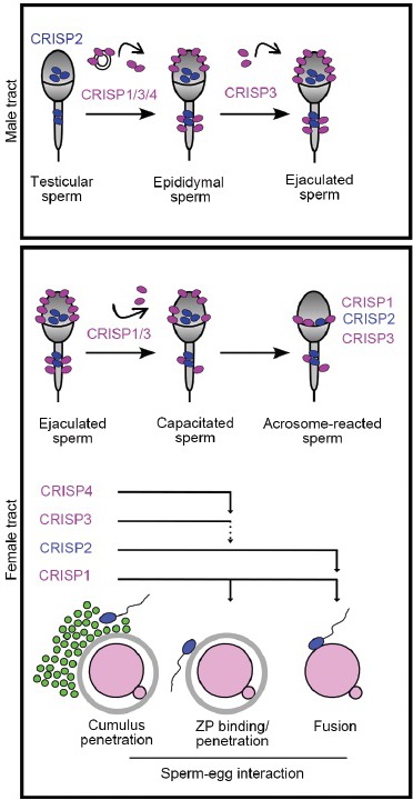
Mammalian fertilization is a complex process that involves different steps of interaction between the male and female gametes. In spite of its relevance, the molecular mechanisms underlying this process still remain to be elucidated. The present review describes the contribution of our laboratory to the understanding of mammalian fertilization using Cysteine-RIch Secretory Proteins (CRISP) as model molecules. Substantial evidence obtained from in vitro assays and knockout models shows that epididymal CRISP1 associates with the sperm surface with two different affinities during maturation, and participates in the regulation of signaling pathways during capacitation as well as in both sperm-zona pellucida interaction and gamete fusion. These observations can be extended to humans as judged by our findings showing that the human homolog of the rodent protein (hCRISP1) is also involved in both stages of fertilization. Evidence supports that other members of the CRISP family secreted in the testis (CRISP2), epididymis (CRISP3-4) or during ejaculation (CRISP3) are also involved in sperm-egg interaction, supporting the existence of a functional redundancy and cooperation between homolog proteins ensuring the success of fertilization. Together, our observations indicate that CRISP proteins accompany spermatozoa along their transit through both the male and female reproductive tracts. We believe these results not only contribute to a better mechanistic understanding of fertilization but also support CRISP proteins as excellent candidates for future research on infertility and contraception.
Figures
Similar articles
-
Epididymal protein CRISP1 plays different roles during the fertilization process.J Androl. 2011 Nov-Dec;32(6):672-8. doi: 10.2164/jandrol.110.012922. Epub 2011 Mar 25. J Androl. 2011. PMID: 21441424 Review.
-
Participation of cysteine-rich secretory proteins (CRISP) in mammalian sperm-egg interaction.Int J Dev Biol. 2008;52(5-6):737-42. doi: 10.1387/ijdb.072538dc. Int J Dev Biol. 2008. PMID: 18649285 Review.
-
Participation of epididymal cysteine-rich secretory proteins in sperm-egg fusion and their potential use for male fertility regulation.Asian J Androl. 2007 Jul;9(4):528-32. doi: 10.1111/j.1745-7262.2007.00283.x. Asian J Androl. 2007. PMID: 17589791 Review.
-
Cysteine-rich secretory proteins (CRISP) and their role in mammalian fertilization.Biol Res. 2011;44(2):135-8. Epub 2011 Sep 20. Biol Res. 2011. PMID: 22513415 Review.
-
Relevance of CRISP proteins for epididymal physiology, fertilization, and fertility.Andrology. 2019 Sep;7(5):610-617. doi: 10.1111/andr.12638. Epub 2019 Jun 19. Andrology. 2019. PMID: 31218833 Review.
Cited by
-
Regulation of Functional Protein Aggregation by Multiple Factors: Implications for the Amyloidogenic Behavior of the CAP Superfamily Proteins.Int J Mol Sci. 2020 Sep 7;21(18):6530. doi: 10.3390/ijms21186530. Int J Mol Sci. 2020. PMID: 32906672 Free PMC article. Review.
-
Role of Zinc (Zn) in Human Reproduction: A Journey from Initial Spermatogenesis to Childbirth.Int J Mol Sci. 2021 Feb 22;22(4):2188. doi: 10.3390/ijms22042188. Int J Mol Sci. 2021. PMID: 33671837 Free PMC article. Review.
-
Expression Analysis of the CRISP2, CATSPER1, PATE1 and SEMG1 in the Sperm of Men with Idiopathic Asthenozoospermia.J Reprod Infertil. 2019 Apr-Jun;20(2):70-75. J Reprod Infertil. 2019. PMID: 31058050 Free PMC article.
-
Detection of Protein Biomarkers Relevant to Sperm Characteristics and Fertility in Semen in Three Wild Felidae: The Flat-Headed Cat (Prionailurus planiceps), Fishing Cat (Prionailurus viverrinus), and Asiatic Golden Cat (Catopuma temminckii).Animals (Basel). 2024 Mar 28;14(7):1027. doi: 10.3390/ani14071027. Animals (Basel). 2024. PMID: 38612267 Free PMC article.
-
Silencing HE4 alleviates the renal fibrosis in lupus nephritis mice by regulating the C3/MMPs/prss axis.Naunyn Schmiedebergs Arch Pharmacol. 2024 Jul;397(7):4823-4831. doi: 10.1007/s00210-023-02883-x. Epub 2023 Dec 29. Naunyn Schmiedebergs Arch Pharmacol. 2024. PMID: 38157023 Free PMC article.
References
-
- Cameo MS, Blaquier J. Androgen-controlled specific proteins in rat epididymis. J Endocrinol. 1976;69:317–24. - PubMed
-
- Garberi JC, Kohane AC, Cameo MS, Blaquier JA. Isolation and characterization of specific rat epididymal proteins. Mol Cell Endocrinol. 1979;13:73–82. - PubMed
-
- Garberi JC, Fontana JD, Blaquier JA. Carbohydrate composition of specific rat epididymal protein. Int J Androl. 1982;5:619–26. - PubMed
-
- Kohane AC, Cameo MS, Piñeiro L, Garberi JC, Blaquier JA. Distribution and site of production of specific proteins in the rat epididymis. Biol Reprod. 1980;23:181–7. - PubMed
-
- Kohane AC, González Echeverría FM, Piñeiro L, Blaquier JA. Interaction of proteins of epididymal origin with spermatozoa. Biol Reprod. 1980;23:737–42. - PubMed
Publication types
MeSH terms
Substances
LinkOut - more resources
Full Text Sources
