Comparative Study
. 2015 Jun 26:12:17.
doi: 10.1186/s12989-015-0094-4.

Acute and subchronic exposure to air particulate matter induces expression of angiotensin and bradykinin-related genes in the lungs and heart: Angiotensin-II type-I receptor as a molecular target of particulate matter exposure

Affiliations
Comparative Study

Acute and subchronic exposure to air particulate matter induces expression of angiotensin and bradykinin-related genes in the lungs and heart: Angiotensin-II type-I receptor as a molecular target of particulate matter exposure

Octavio Gamaliel Aztatzi-Aguilar et al. Part Fibre Toxicol. .

Abstract

Background: Particulate matter (PM) adverse effects on health include lung and heart damage. The renin-angiotensin-aldosterone (RAAS) and kallikrein-kinin (KKS) endocrine systems are involved in the pathophysiology of cardiovascular diseases and have been found to impact lung diseases. The aim of the present study was to evaluate whether PM exposure regulates elements of RAAS and KKS.

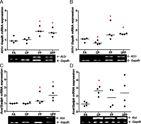
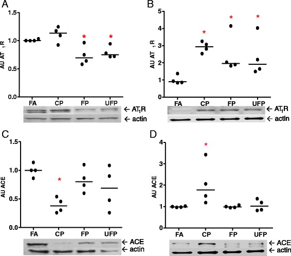
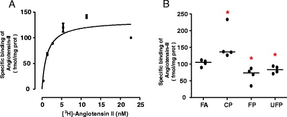
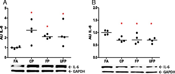
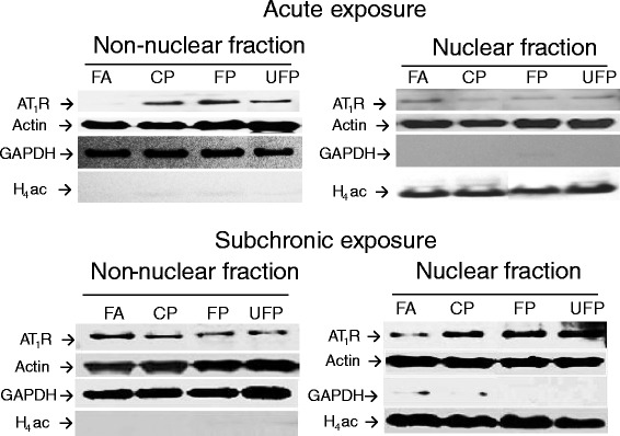
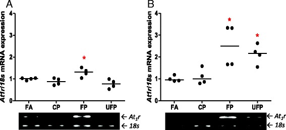
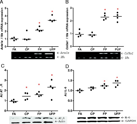
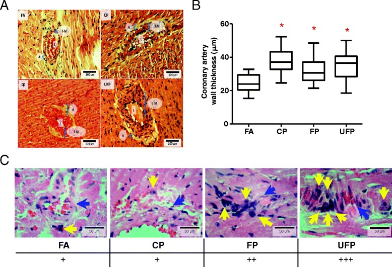
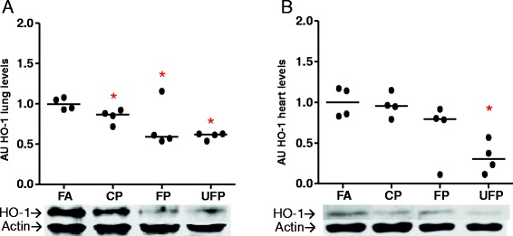
Methods: Sprague-Dawley rats were acutely (3 days) and subchronically (8 weeks) exposed to coarse (CP), fine (FP) or ultrafine (UFP) particulates using a particulate concentrator, and a control group exposed to filtered air (FA). We evaluated the mRNA of the RAAS components At1, At2r and Ace, and of the KKS components B1r, B2r and Klk-1 by RT-PCR in the lungs and heart. The ACE and AT1R protein were evaluated by Western blot, as were HO-1 and γGCSc as indicators of the antioxidant response and IL-6 levels as an inflammation marker. We performed a binding assay to determinate AT1R density in the lung, also the subcellular AT1R distribution in the lungs was evaluated. Finally, we performed a histological analysis of intramyocardial coronary arteries and the expression of markers of heart gene reprogramming (Acta1 and Col3a1).

Results: The PM fractions induced the expression of RAAS and KKS elements in the lungs and heart in a time-dependent manner. CP exposure induced Ace mRNA expression and regulated its protein in the lungs. Acute and subchronic exposure to FP and UFP induced the expression of At1r in the lungs and heart. All PM fractions increased the AT1R protein in a size-dependent manner in the lungs and heart after subchronic exposure. The AT1R lung protein showed a time-dependent change in subcellular distribution. In addition, the presence of AT1R in the heart was accompanied by a decrease in HO-1, which was concomitant with the induction of Acta1 and Col3a1 and the increment of IL-6. Moreover, exposure to all PM fractions increased coronary artery wall thickness.

Conclusion: We demonstrate that exposure to PM induces the expression of RAAS and KKS elements, including AT1R, which was the main target in the lungs and the heart.


Figures

Fig. 1
Fig. 1
Particulate matter induces lung AT1R and ACE mRNA in a size- and time-dependent manner. The animals were exposed to coarse (CP), fine (CP) and ultrafine particulate (UFP). A control group was exposed to filtered air (FA). The semi-quantitative levels of Angiotensin Receptor Type-1 (At1r) and Angiotensin-I Converting Enzyme (Ace) mRNA after acute (3 days, 5 h/d) and subchronic (8 weeks, 5 h/d, 4 d/week) exposure are shown. Scatter dot plot shows the value of the median. Below each graph representatives gels illustrating the expression levels of mRNA are shown. * indicates significant differences among groups (n = 4 per group, p < 0.05)
Fig. 2
Fig. 2
Particle matter exposure regulates AT1R and ACE protein in a particle size-dependent manner. Animals were exposed to coarse (CP), fine (FP) and ultrafine particulate (UFP), or filtered air (FA). a) and b) The protein levels of Angiotensin Receptor Type-1 (AT1R) and c) and d) Angiotensin-I Converting Enzyme (ACE), after acute (3 days, 5 h/day) and subchronic (8 weeks, 5 h/day, 4 days/week) exposure, respectively, are shown as arbitrary units (AU). Scatter dot plot shows the value of the median. * indicates significant differences among groups (n = 4 per group, p < 0.05)
Fig. 3
Fig. 3
Acute exposure to PM modifies [3H]-Angiotensin-II binding to the lung tissue membranes. a) Saturation binding. Membranes, obtained from naïve animals as described in Methods, were incubated with the indicated concentrations of [3H]-Angiotensin-II. Specific receptor binding was determined by subtracting the binding in the presence of 100 μM telmisartan from the total binding. The points show the means from quadruplicate determinations from a single experiment, which was repeated twice more with membranes obtained from different naïve animals. The line drawn is the best fit to a hyperbola. Best-fit values for the equilibrium dissociation constant (K d) and maximum binding (Bmax) are given in the text. b) Single-point determinations. Membranes were obtained from animals exposed to coarse (CP), fine (FP) and ultrafine particulate (UFP) or from the filtered air control group (FA), and then incubated with 10 nM [3H]-Angiotensin-II. Specific receptor binding was determined by subtracting the binding in the presence of 10 μM telmisartan. Scatter dot plot shows the value of the median. * Indicates significant differences among groups (n = 4 per group, p < 0.05)
Fig. 4
Fig. 4
Particle matter modifies the AT1R subcellular distribution in lung tissue in a time-dependent manner. Representative gels of Angiotensin-II type-I receptor (AT1R) detection in lung nuclear and non-nuclear fractions after acute (upper panel) and subchronic (lower panel) exposure to coarse particulate (CP), fine particulate (FP), ultrafine particulate (UFP) or filtered air (AF). We used GAPDH and acetylated Histone-4 (H4ac) as cytosolic and nuclear quality control targets, respectively, and actin was used as a general protein loading control. Representative blot of n = 4
Fig. 5
Fig. 5
Acute, but not subchronic, exposure to particle matter increases IL-6 in lungs. Animals were exposed to coarse (CP), fine (FP) and ultrafine particulate (UFP), or filtered air (FA). The protein interleukine-6 (IL-6) levels after a) acute (3 days, 5 h/day) and b) subchronic exposure (8 weeks, 5 h/day, 4 days/week), are shown as arbitrary units (AU). Scatter dot plot shows the value of the median. * indicates significant differences among groups (n = 4 per group, p < 0.05)
Fig. 6
Fig. 6
Acute and subchronic exposures to fine and ultrafine PM up-regulate heart AT1R mRNA. The animals were acutely (5 h/day, 3 days) or subchronically (5 h/day, 4 days/week, 8 weeks) exposed to coarse (CP), fine (FP) and ultrafine particulate (UFP) or to filtered air (FA). Semi-quantitative levels of Angiotensin Receptor type-1 (At1r) mRNA after a) acute and b) subchronic exposures in the heart. Scatter dot plot shows the value of the median. * indicates significant differences among groups (n = 4 per group, p < 0.05)
Fig. 7
Fig. 7
Subchronic exposure to fine and ultrafine PM induces heart reprogramming and up-regulates AT1R and IL-6. The animals were subchronically exposed (5 h/day, 4 days/week for 8 weeks) to coarse (CP), fine (FP) and ultrafine particulate (UFP) or to filtered air (FA). a) Semi-quantitative levels of alpha-skeletal actin (Acta1a) mRNA and b) Semi-quantitative levels of collagen-III (Col3a1) mRNA in the heart. Protein levels of AT1R and interleukin-6 (IL-6) are expressed in arbitrary units (AU), c) and d), respectively. Scatter dot plot shows the value of the median. * indicates significant differences among groups (n = 4 per group, p < 0.05)
Fig. 8
Fig. 8
Subchronic PM exposure increases intramyocardial coronary artery wall thickness independently of PM size. Hematoxylin & Eosin staining of 5 μm myocardial slices. a) Representative slides of the hearts from rats exposed to Filtered Air (FA), coarse (CP), fine (FP) and ultrafine (UFP) particulates for 5 h/day, 4 days/week for 8 weeks. b) Box plot of ten random measurements of each coronary artery wall measured in the tissues. c) Semi-quantitative analysis of the presence of mononuclear cells in heart coronary arteries in the groups exposed to FP and UFP. L: Artery lumen; A: Tunica adventitia; I-M: Tunica intima-media. Yellow arrows indicate mononuclear cells and blue arrows indicate a blood vessel. Scatter dot plot shows the value of the median. * indicates significant differences among groups (n = 4 per group, p < 0.05)
Fig. 9
Fig. 9
Particle size-dependent decreases in heme oxygenase-1 after subchronic exposure in the lungs and heart. Animals were exposed subchronic to coarse (CP), fine (FP) and ultrafine particulate (UFP) or to filtered air as a control (FA) for 8 weeks (5 h/day, 4 days/week). Protein levels of heme oxygenase-1 (HO-1) in arbitrary units (AU) in lungs a) and heart b). Scatter dot plot shows the value of the median. * indicates significant differences among groups (n = 4 per group, p < 0.05)

References

    1. Pope CA, III, Dockery DW. Health effects of fine particulate air pollution: lines that connect. J Air Waste Manage Assoc. 2006;56:709–742. doi: 10.1080/10473289.2006.10464485. - DOI - PubMed
    1. Simkhovich BZ, Kleinman MT, Kloner RA. Air pollution and cardiovascular injury: epidemiology, toxicology, and mechanisms. J Am Coll Cardiol. 2008;52:719–726. doi: 10.1016/j.jacc.2008.05.029. - DOI - PubMed
    1. Pope CA, Burnett RT, Thurston GD, Thun MJ, Calle EE, Krewski D, et al. Cardiovascular mortality and long-term exposure to particulate air pollution epidemiological evidence of general pathophysiological pathways of disease. Circulation. 2004;109:71–77. doi: 10.1161/01.CIR.0000108927.80044.7F. - DOI - PubMed
    1. Donaldson K, Stone V, Seaton A, MacNee W. Ambient particle inhalation and the cardiovascular system: potential mechanisms. Environ Health Perspect. 2001;109:523. doi: 10.1289/ehp.01109s4523. - DOI - PMC - PubMed
    1. Mutlu GM, Green D, Bellmeyer A, Baker CM, Burgess Z, Rajamannan N, et al. Ambient particulate matter accelerates coagulation via an IL-6–dependent pathway. J Clin Invest. 2007;117:2952–2961. doi: 10.1172/JCI30639. - DOI - PMC - PubMed

Publication types

MeSH terms