Review
. 2015 Nov:49:67-81.
doi: 10.1016/j.preteyeres.2015.06.002. Epub 2015 Jun 23.

Molecular pathogenesis of retinal and choroidal vascular diseases

Affiliations
Review

Molecular pathogenesis of retinal and choroidal vascular diseases

Peter A Campochiaro. Prog Retin Eye Res. 2015 Nov.

Abstract

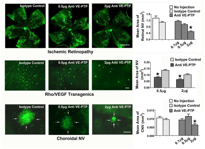
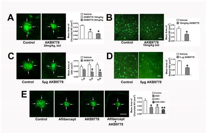
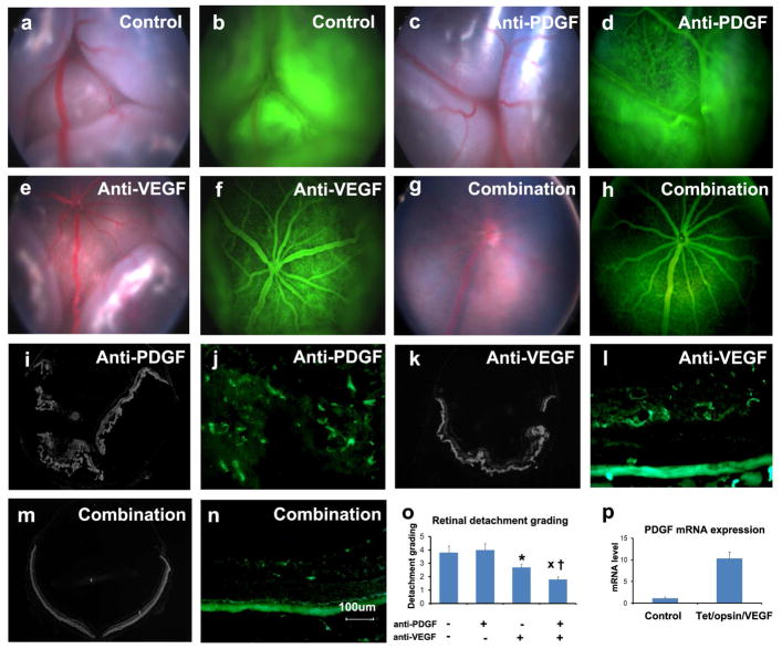
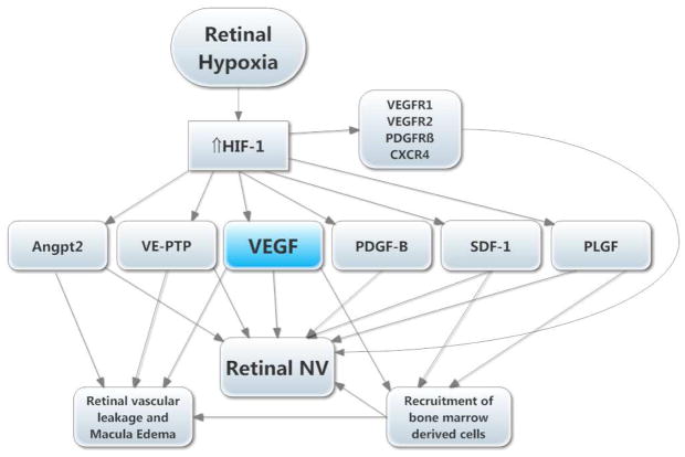
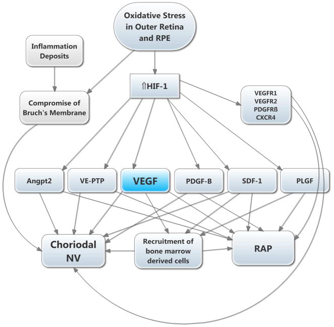
There are two major types of ocular neovascularization that affect the retina, retinal neovascularization (NV) and subretinal or choroidal NV. Retinal NV occurs in a group of diseases referred to as ischemic retinopathies in which damage to retinal vessels results in retinal ischemia. Most prevalent of these are diabetic retinopathy and retinal vein occlusions. Subretinal and choroidal NV occur in diseases of the outer retina and Bruch's membrane, the most prevalent of which is age-related macular degeneration. Numerous studies in mouse models have helped to elucidate the molecular pathogenesis underlying retinal, subretinal, and choroidal NV. There is considerable overlap because the precipitating event in each is stabilization of hypoxia inducible factor-1 (HIF-1) which leads to upregulation of several hypoxia-regulated gene products, including vascular endothelial growth factor (VEGF), angiopoietin 2, vascular endothelial-protein tyrosine phosphatase (VE-PTP), and several others. Stimulation of VEGF signaling and suppression of Tie2 by angiopoietin 2 and VE-PTP are critical for sprouting of retinal, subretinal, and choroidal NV, with perturbation of Bruch's membrane also needed for the latter. Additional HIF-1-regulated gene products cause further stimulation of the NV. It is difficult to model macular edema in animals and therefore proof-of-concept clinical trials were done and demonstrated that VEGF plays a central role and that suppression of Tie2 is also important. Neutralization of VEGF is currently the first line therapy for all of the above disease processes, but new treatments directed at some of the other molecular targets, particularly stabilization of Tie2, are likely to provide additional benefit for subretinal/choroidal NV and macular edema. In addition, the chronicity of these diseases as well as the implication of VEGF as a cause of retinal nonperfusion and progression of background diabetic retinopathy make sustained delivery approaches for VEGF antagonists a priority.

Keywords: Age-related macular degeneration; Angiogenesis; Angiopoietins; Diabetic retinopathy; Hypoxia-inducible factor-1; Platelet-derived growth factor; TIE2; Vascular endothelial growth factor; Vascular endothelial-protein tyrosine phosphatase.


Figures

Figure 1
Figure 1. Schematic showing the vascular supply of the retina
The retinal arteries branch to form the superficial capillary bed near the surface of the retina and send penetrating branches to form the intermediate and deep capillaries. Retinal vessels supply the inner two-thirds of the retina with oxygen and nutrients. The outer third of the retina which consists of photoreceptor outer and inner segments and cells bodies is avascular. It receives oxygen and nutrients from the choroidal circulation. Large choroidal vessels branch and become progressively smaller until they form the choriocapillaris which is fenestrated and allows plasma to pool along Bruch’s membrane. The RPE, which has barrier characteristics prevents fluid from entering the outer retina but allows oxygen and nutrients to enter.
Figure 2
Figure 2. Ocular sections from mouse models of ocular neovascularization (NV)
(A) A normal mouse retina histochemically stained with Griffonia simplicifolia lectin that selectively stains vascular cells and counter-stained to show other retinal cells illustrates the 3 capillary beds of the retina, the superficial capillaries near the surface, the intermediate capillaries at the inner border of the inner nuclear layer, and the deep capillaries at the outer border of the inner nuclear layer. There are also some penetrating vessels showing connections or part of connections between the beds. (B) An ocular section from a mouse with oxygen-induced ischemic retinopathy shows dilation of retinal vessels making it easier to see connections between the three capillary beds. There is retinal NV on the surface of the retina extending into the vitreous cavity (arrows). (C) This is an ocular section from the eye of a rho/VEGF transgenic mouse in which the rhodopsin promoter drives expression of VEGF in photoreceptors. These mice sprout new vessels from the deep capillaries of the retina resulting in new vessels that penetrate through the outer nuclear layer of the retina into the subretinal space. This is a model of retinal angiomatous proliferation which occurs in patients with neovascular age-related macular degeneration. (D) This is an ocular section from a mouse with choroidal NV after laser-induced rupture of Bruch’s membrane. The arrow shows a vessel extending from the choroid through the rupture in Bruch’s membrane and retinal pigmented epithelium into the subretinal space where it connects to a large convoluted network of new vessels, many of which are cut in cross section to show their lumens (astericks). The superior margin of the choroidal NV that borders the photoreceptors is shown by the arrowheads.
Figure 3
Figure 3. Selective staining of neovascularization (NV) in retina of mouse with oxygen-induced ischemic retinopathy
A retinal from a mouse with ischemic retinopathy was stained with FITC-labeled Griffonia simplicifolia lectin for 20 minutes which stains retinal NV (arrowheads) and hyaloid vessels (arrows) but not the pre-existent retinal vessels which facilitates quantification of NV by image analysis. After radial cuts, the retina was flat-mounted allowing visualization of the entire retina and measurement of all retinal NV.
Figure 4
Figure 4. Treatment of Tet/opsin/VEGF double transgenic mice with doxycycline results in exudative retinal detachments that can be imaged by fundus photography
A male Tet/opsin/VEGF double transgenic mouse was given normal drinking water (A) and another was given water containing 2 mg/ml doxycycline in drinking water for 3 days (B). Fundus photography shows a normal retina in the mouse given normal drinking water (A) and an exudative retinal detachment due to severe VEGF-induced vascular leakage in the doxycycline-treated mouse (B). The entire retina is detached giving the retina an opalescent appearance with folds seen superiorly. Also the retinal vessels are dilated giving the vessels a dark red appearance.
Figure 5
Figure 5. Inhibition of vascular endothelial-protein tyrosine phosphatase (VE-PTP) suppresses ocular neovascularization (NV)
At P12, mice with oxygen-induced ischemic retinopathy were given an intravitreous injection in one eye of 0.1, 0.5, or 2 μg of an antibody directed against VE-PTP or 2 μg of IgG isotype control (n≥12 for each). At P17, staining with GSA lectin showed extensive retinal NV (all of the dark green areas are retinal NV) on the surface of the retina in control IgG-injected eyes and significantly less in eyes injected with 2 μg of anti-VE-PTP (top row, *p<0.001 for comparison with IgG control by one-way ANOVA and Bonferroni to adjust for multiple comparisons; bar=500μm). At P15, rho/VEGF transgenic mice were given an intravitreous injection of 0.5 or 2 μg of anti-VE-PTP in one eye and a corresponding dose of control IgG in the fellow eye (n=6 for each). At P21, retinas were stained with GSA lectin and flat mounted with photoreceptor side facing up. All of the dark green spots are subretinal NV and there was significantly less in eyes injected with 0.5 or 2 μg of anti-VE-PTP than corresponding IgG control eyes (middle row, *p=0.01 by unpaired t-test for comparison with fellow eye IgG control; bar=100μm). After laser-induced rupture of Bruch’s membrane in each eye, mice had intravitreous injection of 0.1, 0.5 or 2 μg of anti-VE-PTP in one eye and 2 μg of control IgG or no injection in the other eye (n≥12 for each). After 7 days, eyecups were stained with GSA lectin and flat mounted. The area of choroidal NV was significantly less in eyes injected with 2 μg of anti-VE-PTP compared with control IgG (bottom row, *p<0.001 by one-way ANOVA and Bonferroni, bar=100μm).
Figure 6
Figure 6. Subcutaneous or intraocular injection of AKB-9778 suppresses choroidal and subretinal neovascularization (NV)
After laser-induced rupture of Bruch’s membrane at 3 locations in each eye, mice were given subcutaneous injections of vehicle or 10 or 20 mg/kg AKB-9778 (n=10 for each) twice a day or a single intraocular injection of 1, 3, or 5 μg of AKB9778 in one eye and vehicle in the fellow eye (n=10 for each group). After 7 days, eyecups were stained with FITC-labeled Griffonia simplicifolia (GSA) lectin. Starting at P15, hemizygous rho/VEGF transgenic mice were injected subcutaneously with AKB-9778 (10 mg/kg bid) or vehicle (n=10 for each) or given an intraocular injection of 3 μg of AKB-9778 in one eye and vehicle in the fellow eye (n=10). At P21, retinas were stained with GSA lectin and retinas were flat-mounted. (A) Subcutaneous injection of 10 or 20 mg/kg of AKB-9778 caused significant reduction in choroidal NV (*p=0.03;**p=0.004 for comparison with control by one-way ANOVA and Bonferroni to adjust for multiple comparisons, bar=100μm). (B) Subcutaneous injection of 10 mg/kg of AKB-9778 significantly reduced the area of subretinal NV in rho/VEGF transgenics (p=0.038 for comparison with control by unpaired t-test, bar=100μm). (C) Intraocular injection of 3 or 5 μg, but not 1 μg of AKB-9778 significantly reduced the area of choroidal NV (*p<0.01 for comparison with vehicle control by one-way ANOVA and Bonferroni, bar=100μm). (D) Intraocular injection of 5 μg of AKB-9778 significantly reduced subretinal NV in rho/VEGF transgenic mice (p=0.022 for difference from vehicle control by unpaired t-test, bar=100μm). In an independent experiment, mice had rupture of Bruch’s membrane with laser and then had intraocular injection of 40 μg of the anti-VEGF agent aflibercept or PBS and subcutaneous injections of 20 mg/kg AKB-9778 or PBS twice a day. This resulted in 4 groups of mice with n=19 in each group: control, aflibercept, AKB-9778, or aflibercept + AKB-9778. After 7 days, compared with the area of GSA lectin-stained choroidal NV in control mice, there was a significant reduction in aflibercept- and AKB-9778- treated mice (E, *p<0.01 by ANOVA with Dunnett’s correction). Compared with mice treated with aflibercept or AKB-9778, those treated with the combination had significantly less choroidal NV (**p<0.05 by ANOVA with Dunnett’s correction).
Figure 7
Figure 7. Prevention of exudative retinal detachment in Tet/opsin/VEGF double transgenic mice is greater with both anti-PDGF-BB designed ankyrin repeat protein (DARPin) and anti-VEGF-A DARPin than either alone
Adult male Tet/opsin/VEGF double transgenic mice were given daily subcutaneously injections of 50mg/kg of doxcycline and intraperitoneal injections of PBS as control (a,b), 1mg/kg anti-PDGF-BB DARPin (c,d), 1mg/kg anti-VEGF-A DARPin (e,f), or 1mg/kg of both DARPins (g,h). After 4 days, fundus photographs and fluorescein angiograms showed total bullous retinal detachments in eyes of mice treated with PBS (a,b) or anti-PDGF-BB DARPin (c,d), partial retinal detachments in eyes of mice treated with anti-VEGF-A DARPin (e,f), and little or no retinal detachment in eyes of mice treated with both DARPins (g,h). Ocular sections stained with Hoechst or FITC-Griffonia simplicifolia lectin confirmed total retinal detachments (i) and extensive NV thoughout the outer retina (j) in eyes of mice treated with anti-PDGF-BB DARPin, partial retinal detachments (k) and dilated vessels with NV extending from deep capillaries (l) in eyes of mice treated with anti-VEGF-A DARPin, and no retinal detachments (M) and normal vessels with little or no NV (n) in eyes of mice treated with both DARPins. Grading of the incidence and severity of leakage or retinal detachments (o) showed that eyes from mice treated with both DARPins had significantly less severe grades than those seen in eyes of mice treated with anti-VEGF-A DARPin (x, p=0.0329) or anti-PDGF-BB DARPin (†, p=0.0098) by Wilcoxin test. (p) Tet/Opsin/VEGF mice and C57BL/6 mice (n=5 for each) were treated with doxycycline and after 3 days, the relative of expression of Pdgfb mRNA normalized to cyclophilin A mRNA was significantly greater in Tet/Opsin/VEGF mice (p=0.0002 by unpaired t-test).
Figure 8
Figure 8. Molecular pathogenesis of retinal neovascularization (NV)
A simplified version of the molecular pathogenesis of retinal NV illustrates several important molecular signals. It highlights soluble mediators and omits cell-cell and cell-matrix signaling. Retinal NV occurs in diabetic retinopathy and other ischemic retinopathies. The underlying disease process (e.g. high glucose in diabetic retinopathy) damages retinal vessels causing vessel closure and retinal ischemia, which results in elevated HIF-1 levels. HIF-1 upregulates several vasoactive gene products including angiopoietin 2 (Angpt2), vascular endothelial-protein tyrosine phosphatase (VE-PTP), vascular endothelial growth factor (VEGF), platelet-derived growth factor (PDGF-B), stromal-derived growth factor (SDF-1), placental growth factor (PLGF), and several of their receptors. VEGF causes vascular leakage and in combination with Angpt2 and VE-PTP causes sprouting of new vessels. VEGF, SDF-1, and PLGF recruit bone marrow-derived cells which provide paracrine stimulation. PDGF-B recruits pericytes which also provide paracrine stimulation.
Figure 9
Figure 9. Molecular pathogenesis of subretinal neovascularization (NV)
This schematic highlights several important molecular signals involved in subretinal NV. Oxidative stress in the retinal pigmented epithelium (RPE) and photoreceptors causes increased levels of HIF-1, which upregulates vasoactive gene products as described above. Retinal angiomatous proliferation (RAP) occurs if VEGF levels in photoreceptors are sufficiently high to cause a gradient that reaches the deep capillary bed of the retina where there is constitutive expression of Angpt2. Choroidal NV occurs if there is elevation of VEGF and Angpt2 combined with perturbation of Bruch’s membrane and the RPE. The other HIF-1-responsive gene products fuel the process similar to the situation in retinal NV.

References

    1. Age-Related Eye Disease Study Research Group. A randomized, placebo-controlled, clinical trial of high-dose supplementation with vitamins C and E, beta carotene, and zinc for age-related macular degeneration and vision loss. Arch Ophthalmol. 2001;119 (10):1417–1436. - PMC - PubMed
    1. Aiello LP, Pierce EA, Foley ED, Takagi H, Chen H, Riddle L, Ferrara N, King GL, Smith LEH. Suppression of retinal neovascularization in vivo by inhibition of vascular endothelial growth factor (VEGF) using soluble VEGF-receptor chimeric proteins. Proc Natl Acad Sci USA. 1995;92 (23):10457–10461. - PMC - PubMed
    1. Alon T, Hemo I, Itin A, Pe’er J, Stone J, Keshet E. Vascular endothelial growth factor acts as a survival factor for newly formed retinal vessels and has implications for retinopathy of prematurity. Nature Med. 1995;1 (10):1024–1028. - PubMed
    1. Auricchio A, Behling KC, Maguire AM, O’Conner EM, Bennett J, Wilson JM, Tolentino MJ. Inhibition of retinal neovascularization by intraocular viral-mediated delivery of anti-angiogenic agents. Mol Ther. 2002;6 (4):490–494. - PubMed
    1. Avery RL, Castellarin AA, Steinle NC, Dhoot DS, Pieramici DJ, See R, Couvillion S, Nasir MA, Rabena MD, Le K, Maia M, Visich JE. Systemic pharmacokinenetics following intravitreal injections of ranibizumab, bevacizumab or aflibercept in patients with neovascular AMD. Br J Ophthalmol. 2014;98 (12):1543–1546. - PMC - PubMed

Publication types

MeSH terms

Substances