

Osteoporosis imaging: effects of bone preservation on MDCT-based trabecular bone microstructure parameters and finite element models
- PMID: 26113362
- PMCID: PMC4482285
- DOI: 10.1186/s12880-015-0066-z
Osteoporosis imaging: effects of bone preservation on MDCT-based trabecular bone microstructure parameters and finite element models
Abstract
Background: Osteoporosis is defined as a skeletal disorder characterized by compromised bone strength due to a reduction of bone mass and deterioration of bone microstructure predisposing an individual to an increased risk of fracture. Trabecular bone microstructure analysis and finite element models (FEM) have shown to improve the prediction of bone strength beyond bone mineral density (BMD) measurements. These computational methods have been developed and validated in specimens preserved in formalin solution or by freezing. However, little is known about the effects of preservation on trabecular bone microstructure and FEM. The purpose of this observational study was to investigate the effects of preservation on trabecular bone microstructure and FEM in human vertebrae.
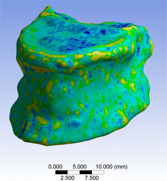
Methods: Four thoracic vertebrae were harvested from each of three fresh human cadavers (n=12). Multi-detector computed tomography (MDCT) images were obtained at baseline, 3 and 6 month follow-up. In the intervals between MDCT imaging, two vertebrae from each donor were formalin-fixed and frozen, respectively. BMD, trabecular bone microstructure parameters (histomorphometry and fractal dimension), and FEM-based apparent compressive modulus (ACM) were determined in the MDCT images and validated by mechanical testing to failure of the vertebrae after 6 months.
Results: Changes of BMD, trabecular bone microstructure parameters, and FEM-based ACM in formalin-fixed and frozen vertebrae over 6 months ranged between 1.0-5.6% and 1.3-6.1%, respectively, and were not statistically significant (p>0.05). BMD, trabecular bone microstructure parameters, and FEM-based ACM as assessed at baseline, 3 and 6 month follow-up correlated significantly with mechanically determined failure load (r=0.89-0.99; p<0.05). The correlation coefficients r were not significantly different for the two preservation methods (p>0.05).
Conclusions: Formalin fixation and freezing up to six months showed no significant effects on trabecular bone microstructure and FEM-based ACM in human vertebrae and may both be used in corresponding in-vitro experiments in the context of osteoporosis.
Figures

Similar articles
-
Prediction of bone strength by μCT and MDCT-based finite-element-models: how much spatial resolution is needed?Eur J Radiol. 2014 Jan;83(1):e36-42. doi: 10.1016/j.ejrad.2013.10.024. Epub 2013 Nov 8. Eur J Radiol. 2014. PMID: 24274992
-
[The texture-analysis of high-resolution computed tomograms as an additional procedure in osteoporosis diagnosis: in-vitro studies on vertebral segments].Rofo. 1999 Aug;171(2):136-42. doi: 10.1055/s-1999-242. Rofo. 1999. PMID: 10506888 German.
-
[Multislice-CT for structure analysis of trabecular bone - a comparison with micro-CT and biomechanical strength].Rofo. 2004 May;176(5):709-18. doi: 10.1055/s-2004-813078. Rofo. 2004. PMID: 15122470 German.
-
Three-dimensional microimaging (MRmicroI and microCT), finite element modeling, and rapid prototyping provide unique insights into bone architecture in osteoporosis.Anat Rec. 2001 Apr;265(2):101-10. doi: 10.1002/ar.1060. Anat Rec. 2001. PMID: 11323772 Review.
-
Imaging of trabecular bone structure in osteoporosis.Eur Radiol. 1999;9(9):1781-8. doi: 10.1007/s003300050922. Eur Radiol. 1999. PMID: 10602950 Review.
Cited by
-
Does formalin fixation influence MSCT/CBCT accuracy?Surg Radiol Anat. 2018 Jan;40(1):31-37. doi: 10.1007/s00276-017-1908-x. Epub 2017 Aug 21. Surg Radiol Anat. 2018. PMID: 28828519
-
Effect of radiation dose reduction on texture measures of trabecular bone microstructure: an in vitro study.J Bone Miner Metab. 2018 May;36(3):323-335. doi: 10.1007/s00774-017-0836-5. Epub 2017 Apr 7. J Bone Miner Metab. 2018. PMID: 28389933
-
Reliability of 3D image analysis and influence of contrast medium administration on measurement of Hounsfield unit values of the proximal femur.PLoS One. 2020 Oct 21;15(10):e0241012. doi: 10.1371/journal.pone.0241012. eCollection 2020. PLoS One. 2020. PMID: 33085702 Free PMC article.
-
Effects of dose reduction on bone strength prediction using finite element analysis.Sci Rep. 2016 Dec 9;6:38441. doi: 10.1038/srep38441. Sci Rep. 2016. PMID: 27934902 Free PMC article.
-
A Comparative Analysis of International Classification Systems to Predict the Risk of Collapse in Single-Level Osteoporotic Vertebral Fractures.Diagnostics (Basel). 2024 Sep 27;14(19):2152. doi: 10.3390/diagnostics14192152. Diagnostics (Basel). 2024. PMID: 39410556 Free PMC article.
References
-
- NIH NIH Consensus Development Panel on Osteoporosis Prevention, Diagnosis, and Therapy, March 7–29, 2000: highlights of the conference. South Med J. 2001;94:569–73. - PubMed
-
- WHO Study Group Assessment of fracture risk and its application to screening for postmenopausal osteoporosis. Report of a WHO Study Group. World Health Organ Tech Rep Ser. 1994;843:1–129. - PubMed
Publication types
MeSH terms
LinkOut - more resources
Full Text Sources
Other Literature Sources
Medical
