. 2015 Jul 17;349(6245):324-8.
doi: 10.1126/science.1260031. Epub 2015 Jun 25.

Circadian rhythms. A protein fold switch joins the circadian oscillator to clock output in cyanobacteria

Affiliations

Circadian rhythms. A protein fold switch joins the circadian oscillator to clock output in cyanobacteria

Yong-Gang Chang et al. Science. .

Abstract

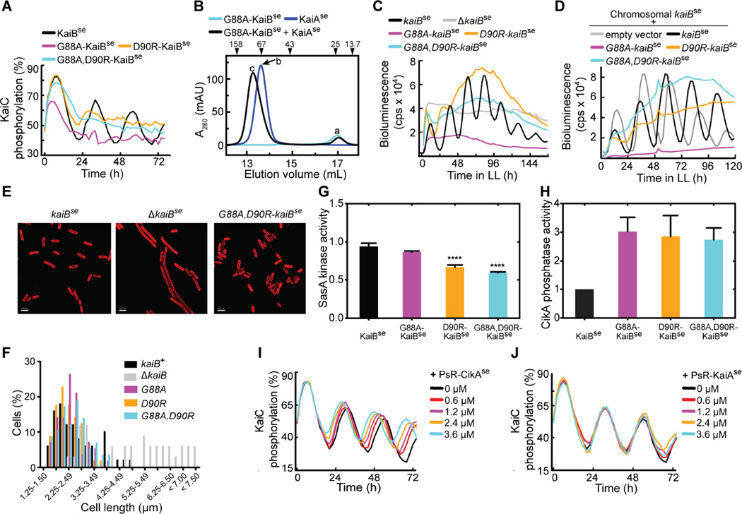
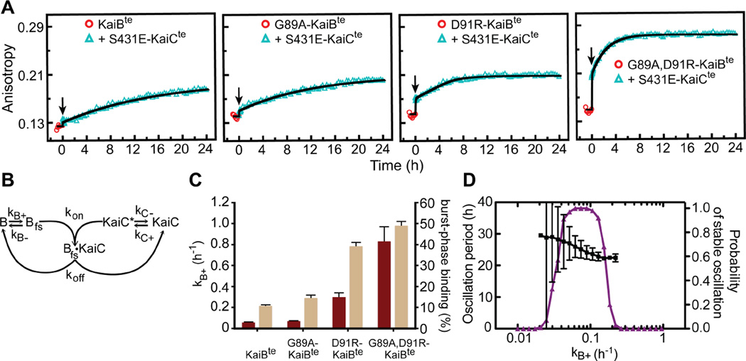
Organisms are adapted to the relentless cycles of day and night, because they evolved timekeeping systems called circadian clocks, which regulate biological activities with ~24-hour rhythms. The clock of cyanobacteria is driven by a three-protein oscillator composed of KaiA, KaiB, and KaiC, which together generate a circadian rhythm of KaiC phosphorylation. We show that KaiB flips between two distinct three-dimensional folds, and its rare transition to an active state provides a time delay that is required to match the timing of the oscillator to that of Earth's rotation. Once KaiB switches folds, it binds phosphorylated KaiC and captures KaiA, which initiates a phase transition of the circadian cycle, and it regulates components of the clock-output pathway, which provides the link that joins the timekeeping and signaling functions of the oscillator.


Figures

Fig. 1
Fig. 1. KaiB switches its fold to bind KaiC
(A) Plots of chemical-shift based secondary structures of KaiBte*, KaiBte* + CIte*, and G89A, D91R-KaiBte* determined by TALOS+ (17). Unassigned proline and non-proline residues are indicated by small red and blue dots along the horizontal axis at y=0. The secondary structures of KaiBte and G89A, D91R-KaiBte*, are shown for comparison. KaiBte* residues Q52 – E56 in the KaiBte*–CIte* complex were not assignable, probably due to exchange broadening. Vertical dashed lines are visual guides separating the N terminal and C-terminal halves of KaiB. (B) Structural comparisons of KaiBte, G89A, D91R-KaiBte*, and N-SasAse. Residues K58, G89 and D91 are highlighted for their roles in fold switching.
Figure 2
Figure 2. KaiB fold switching regulates oscillator function and clock output
(A) In vitro KaiC phosphorylation assays using KaiCse, KaiAse, and KaiBse, G88A-KaiBse, D90R-KaiBse, or G88A, D90R-KaiBse. (B) Gel-filtration profiles of G88A-KaiBse, KaiAse, and G88A-KaiBse + KaiAse. Peaks a–c were analyzed by SDS-PAGE (fig. S13). (C) Bioluminescence from strains that carry a PkaiB luc reporter for circadian rhythmicity. Cells harbored kaiBse, G88AkaiBse, D90R-kaiBse, or G88A, D90R-kaiBse, or cells with kaiBse deletion. (D) Bioluminescence from strains that carry a PkaiB luc reporter expressing kaiBse, G88A-kaiBse, D90R-kaiBse, G88A, D90R-kaiBse, or empty vector, in addition to chromosomal kaiBse. (E) Representative micrographs of cells expressing kaiBse, lacking kaiBse, or harboring G88A, D90R-kaiBse. Cellular autofluorescence in red, scale bars=2.5 microns. (F) Histograms showing cell-length distributions of strains expressing kaiBse, ΔkaiBse, G88A-kaiBse, D90R-kaiBse, or G88A, D90R kaiBse as the only copy of kaiB. (G) SasAse kinase activities in the presence of S431E-KaiCse and KaiBse, G88A-KaiBse, D90R-KaiBse, or G88A, D90R-KaiBse. The mixtures were incubated for 2 hours before SasAse, RpaAse and [γ-32P]-labeled ATP were added. Relative kinase activities compare the mean steady-state amount of 32P-labeled RpaAse to that of a reaction of S431EKaiCse alone (n = 4, error bars denote SEM). One-way ANOVA gives p-value < 0.001, and **** denotes Bonferroni corrected p-values < 0.001 for pairwise comparisons against kinase activity with KaiBse (α = 0.05). (H) CikA phosphatase activity toward phosphorylated RpaA in the presence of KaiCse and KaiBse, G88A-KaiBse, D90R-KaiBse, or G88A, D90R-KaiBse (n = 4–5, error bars denote SEM). KaiCse alone or KaiBse alone did not activate CikA phosphatase activity (fig. S17). (I) In vitro KaiCse phosphorylation assays as a function of concentration of PsRCikAse. (J) Same as (I) except for using PsR-KaiAse instead of PsR-CikAse. The black curves in (I) and (J) are identical.
Figure 3
Figure 3. KaiB fold switching regulates slow formation of the KaiB-KaiC complex
(A) Fluorescence anisotropies of 6-IAF (6-iodoacetamidofluorescein)-labeled KaiBte, G89AKaiBte, D91R-KaiBte, and G89A, D91R-KaiBte in the presence of S431E-KaiCte. KaiB samples were incubated for 1-h (circles) before addition (arrow) of S431E-KaiCte. A54C mutation was introduced to all KaiB for fluorescence labeling. (B) Scheme for modeling. (C) Forward fold-switching rate constants, kB+ (maroon), and burst-phase binding to S431E-KaiCte (tan). Burst-phase binding, defined as the percentage of KaiBte-S431E-KaiCte complexes formed at t = 0.1 h in the model relative to steady state binding at t = 24 h, were derived from fitting data after adding S431E-KaiCte in (A), to the model shown in (B). Burst-phase error bars show the standard deviation from model calculations by bootstrap resampling the raw data (n = 20). kB+ values used in these fits were pre-determined from analysis of the kinetics of binding of KaiBte variants to the isolated CIte* domain (fig. S21), a condition where we assumed the rate-limiting step in complex formation is due only to KaiB fold switching. Error bars for kB+ were estimated by bootstrap resampling the original dataset 500 times. (D) Mathematical modeling of KaiC phosphorylation period (black) and probability of stable oscillation (purple) as a function of, kB+. The black bars indicate the standard deviation in the model period from 100 oscillator calculations at each value of kB+, with the other parameters randomly varied as described in Supplementary Materials.
Figure 4
Figure 4. KaiB and SasA bind to similar sites on CI
(A) EPR-restrained model of the CIte*-N-SasAte complex. The HADDOCK model of the complex with the best score is superimposed on the crystal structure of KaiCte (PDB ID: 4o0m). (B) Qualitative structural model of the interaction of CIte* and fsKaiB (G89A, D91R-KaiBte*), based on HDX-MS data and mutagenesis. Dark blue and cyan spheres represent CI residues whose mutations strongly or moderately weaken binding, respectively. Dark blue and cyan ribbons represent protection against H/D exchange upon complex formation that are >1.5 and 0.5 – 1.5 standard deviations above the average, respectively, as determined by HDX-MS (figs. S32–S35).
Figure 5
Figure 5. Model of KaiB fold switching as linchpin for the cyanobacterial clock
Excursion of KaiB to the rare fold-switch state causes fsKaiB to displace SasA for binding to KaiC. KaiC-stabilized fsKaiB captures KaiA, initiating the dephosphorylation phase of the cycle. These aspects control oscillator period. CikA and KaiA compete for binding to fsKaiB, further linking oscillator function related to KaiA and output activity via CikA-mediated dephosphorylation of RpaA. The competitive interactions of fsKaiB with SasA, and KaiA with CikA, implicate “output components” CikA and SasA as parts of an extended oscillator.

References

    1. Dunlap JC. Molecular basis for circadian clocks. Cell. 1999;96:271–290. - PubMed
    1. Nakajima M, et al. Reconstitution of circadian oscillation of cyanobacterial KaiC phosphorylation in vitro. Science. 2005;308:414–415. - PubMed
    1. Ishiura M, et al. Expression of a gene cluster kaiABC as a circadian feedback process in cyanobacteria. Science. 1998;281:1519–1523. - PubMed
    1. Iwasaki H, Nishiwaki T, Kitayama Y, Nakajima M, Kondo T. KaiA-stimulated KaiC phosphorylation in circadian timing loops in cyanobacteria. Proc. Natl. Acad. Sci. USA. 2002;99:15788–15793. - PMC - PubMed
    1. Williams SB, Vakonakis I, Golden SS, LiWang A. Structure and function from the circadian clock protein KaiA of Synechococcus elongatus: A potential clock input mechanism. Proc. Natl. Acad. Sci. USA. 2002;99:15357–15362. - PMC - PubMed

Publication types

MeSH terms

Substances