. 2015 May 6;6(6):1977-89.
doi: 10.1364/BOE.6.001977. eCollection 2015 Jun 1.

Accessing to arteriovenous blood flow dynamics response using combined laser speckle contrast imaging and skin optical clearing

Affiliations

Accessing to arteriovenous blood flow dynamics response using combined laser speckle contrast imaging and skin optical clearing

Rui Shi et al. Biomed Opt Express. .

Abstract

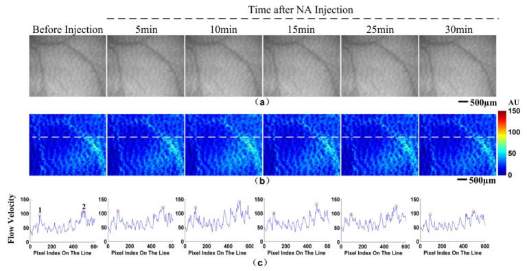
Laser speckle contrast imaging (LSCI) shows a great potential for monitoring blood flow, but the spatial resolution suffers from the scattering of tissue. Here, we demonstrate the capability of a combination method of LSCI and skin optical clearing to describe in detail the dynamic response of cutaneous vasculature to vasoactive noradrenaline injection. Moreover, the superior resolution, contrast and sensitivity make it possible to rebuild arteries-veins separation and quantitatively assess the blood flow dynamical changes in terms of flow velocity and vascular diameter at single artery or vein level.

Keywords: (120.6150) Speckle imaging; (150.1135) Algorithms; (170.1470) Blood or tissue constituent monitoring; (290.0290) Scattering.


Figures

Fig. 1
Fig. 1
The schematic of the LSCI system.
Fig. 2
Fig. 2
Monitoring cutaneous vasculature and blood flow at status of turbid skin; OCA-5 min; and 5-, 10-, 15-, 25-, 30-min after saline injection accompanied with OCA-treatment: white-light images (a); blood flow velocity maps (b); profiles of the flow velocity along the horizontal white line (c), respectively. Bar = 500 μm.
Fig. 3
Fig. 3
The steps of arteries-veins separation (a): speckle contrast image (step 1); vasculature network image (step 2), bright is the vasculature network and dark is non-vascular region; weighted temporal minimum intensity image (step 3); arteries-veins separation image (step 4). Saline injection-induced relative changes in vascular diameter and flow velocity in artery A (b, c); and vein V (d, e) during OCA- treatment. The injection time is set to be 0, which is consistent with the arrow shown in (b-e). Bar = 500 μm.
Fig. 4
Fig. 4
Monitoring cutaneous blood flow dynamics at status of turbid skin; OCA-5 min; and 5-, 10-, 15-, 25-, 30-min after NA injection accompanied with OCA-treatment: white-light images (a); blood flow velocity maps (b); profiles of the flow velocity along the horizontal white line (c), respectively. Bar = 500 μm.
Fig. 5
Fig. 5
Speckle contrast image (a); arteries-veins separation image (b); NA injection-induced relative changes in vascular diameter and flow velocity in artery A1 (c, d); vein V1 (e, f); and vein V2 (g, h) accompanied with OCA- treatment. The injection time is set to be 0, which is consistent with the arrow shown in (c-h). Bar = 500 μm.
Fig. 6
Fig. 6
Monitoring cutaneous blood flow dynamics before NA injection; and 5-, 10-, 15-, 25-, 30-min after NA injection through the turbid skin: white-light images (a); blood flow velocity maps (b); profiles of the flow velocity along the horizontal white line (c), respectively. Bar = 500 μm.

References

    1. Holowatz L. A., Thompson-Torgerson C. S., Kenney W. L., “The human cutaneous circulation as a model of generalized microvascular function,” J. Appl. Physiol. 105(1), 370–372 (2008).10.1152/japplphysiol.00858.2007 - DOI - PubMed
    1. Struijker-Boudier H. A., Rosei A. E., Bruneval P., Camici P. G., Christ F., Henrion D., Lévy B. I., Pries A., Vanoverschelde J. L., “Evaluation of the microcirculation in hypertension and cardiovascular disease,” Eur. Heart J. 28(23), 2834–2840 (2007).10.1093/eurheartj/ehm448 - DOI - PubMed
    1. Quondamatteo F., “Skin and diabetes mellitus: what do we know?” Cell Tissue Res. 355(1), 1–21 (2014).10.1007/s00441-013-1751-2 - DOI - PubMed
    1. Rossi M., Carpi A., Galetta F., Franzoni F., Santoro G., “The investigation of skin blood flowmotion: a new approach to study the microcirculatory impairment in vascular diseases?” Biomed. Pharmacother. 60(8), 437–442 (2006).10.1016/j.biopha.2006.07.012 - DOI - PubMed
    1. Hodges G. J., Del Pozzi A. T., “Noninvasive examination of endothelial, sympathetic, and myogenic contributions to regional differences in the human cutaneous microcirculation,” Microvasc. Res. 93, 87–91 (2014).10.1016/j.mvr.2014.04.002 - DOI - PubMed

LinkOut - more resources