

Phenformin Induces Cell Cycle Change, Apoptosis, and Mesenchymal-Epithelial Transition and Regulates the AMPK/mTOR/p70s6k and MAPK/ERK Pathways in Breast Cancer Cells
- PMID: 26114294
- PMCID: PMC4482683
- DOI: 10.1371/journal.pone.0131207
Phenformin Induces Cell Cycle Change, Apoptosis, and Mesenchymal-Epithelial Transition and Regulates the AMPK/mTOR/p70s6k and MAPK/ERK Pathways in Breast Cancer Cells
Abstract
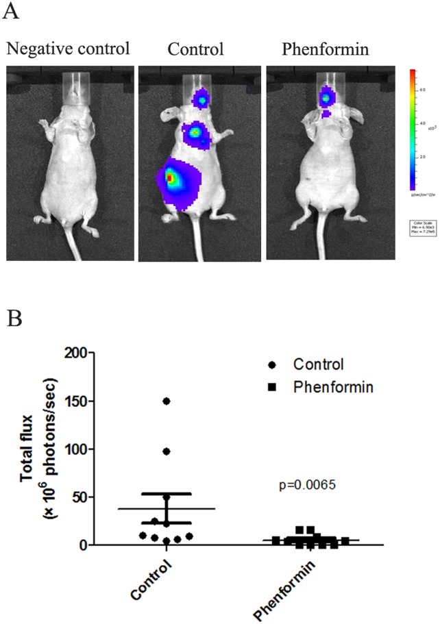
Breast cancer remains a world-wide challenge, and additional anti-cancer therapies are still urgently needed. Emerging evidence has demonstrated the potent anti-tumor effect of biguanides, among which phenformin was reported to potentially be a more active anti-cancer agent than metformin. However, little attention has been given to the role of phenformin in breast cancer. In this study, we reveal the role of phenformin in cell death of the MCF7, ZR-75-1, MDA-MB-231 and SUM1315 breast cancer cell lines. The respective IC50 values of phenformin in MCF7, ZR-75-1, MDA-MB-231 and SUM1315 cells were 1.184±0.045 mM, 0.665±0.007 mM, 2.347±0.010 mM and 1.885±0.015 mM (mean± standard error). Phenformin induced cell cycle change and apoptosis in breast cancer cells via the AMPK/mTOR/p70s6k and MAPK/ERK pathways. Interestingly, phenformin induced MET (mesenchymal-epithelial transition) and decreased the migration rate in breast cancer cell lines. Furthermore, our results suggest that phenformin inhibits breast cancer cell metastasis after intracardiac injection into nude mice. Taken together, our study further confirms the potential benefit of phenformin in breast cancer treatment and provides novel mechanistic insight into its anti-cancer activity in breast cancer.
Conflict of interest statement
Figures


Similar articles
-
The investigational Aurora kinase A inhibitor alisertib (MLN8237) induces cell cycle G2/M arrest, apoptosis, and autophagy via p38 MAPK and Akt/mTOR signaling pathways in human breast cancer cells.Drug Des Devel Ther. 2015 Mar 16;9:1627-52. doi: 10.2147/DDDT.S75378. eCollection 2015. Drug Des Devel Ther. 2015. PMID: 25834401 Free PMC article.
-
The pan-inhibitor of Aurora kinases danusertib induces apoptosis and autophagy and suppresses epithelial-to-mesenchymal transition in human breast cancer cells.Drug Des Devel Ther. 2015 Feb 17;9:1027-62. doi: 10.2147/DDDT.S74412. eCollection 2015. Drug Des Devel Ther. 2015. PMID: 25733818 Free PMC article.
-
Eriocalyxin B, a novel autophagy inducer, exerts anti-tumor activity through the suppression of Akt/mTOR/p70S6K signaling pathway in breast cancer.Biochem Pharmacol. 2017 Oct 15;142:58-70. doi: 10.1016/j.bcp.2017.06.133. Epub 2017 Jun 30. Biochem Pharmacol. 2017. PMID: 28669564
-
Phenformin as an Anticancer Agent: Challenges and Prospects.Int J Mol Sci. 2019 Jul 5;20(13):3316. doi: 10.3390/ijms20133316. Int J Mol Sci. 2019. PMID: 31284513 Free PMC article. Review.
-
Progress in antitumor mechanisms and applications of phenformin (Review).Oncol Rep. 2024 Nov;52(5):151. doi: 10.3892/or.2024.8810. Epub 2024 Sep 20. Oncol Rep. 2024. PMID: 39301645 Free PMC article. Review.
Cited by
-
AMPK in Intestinal Health and Disease: A Multifaceted Therapeutic Target for Metabolic and Inflammatory Disorders.Drug Des Devel Ther. 2025 Apr 21;19:3029-3058. doi: 10.2147/DDDT.S507489. eCollection 2025. Drug Des Devel Ther. 2025. PMID: 40291159 Free PMC article. Review.
-
Mechanism of antineoplastic activity of lonidamine.Biochim Biophys Acta. 2016 Dec;1866(2):151-162. doi: 10.1016/j.bbcan.2016.08.001. Epub 2016 Aug 4. Biochim Biophys Acta. 2016. PMID: 27497601 Free PMC article. Review.
-
AMP-activated protein kinase and energy balance in breast cancer.Am J Transl Res. 2017 Feb 15;9(2):197-213. eCollection 2017. Am J Transl Res. 2017. PMID: 28337254 Free PMC article. Review.
-
Targeting oncogenic KRAS in non-small cell lung cancer cells by phenformin inhibits growth and angiogenesis.Am J Cancer Res. 2015 Oct 15;5(11):3339-49. eCollection 2015. Am J Cancer Res. 2015. PMID: 26807315 Free PMC article.
-
Antidiabetic Biguanides Radiosensitize Hypoxic Colorectal Cancer Cells Through a Decrease in Oxygen Consumption.Front Pharmacol. 2018 Oct 3;9:1073. doi: 10.3389/fphar.2018.01073. eCollection 2018. Front Pharmacol. 2018. PMID: 30337872 Free PMC article.
References
Publication types
MeSH terms
Substances
LinkOut - more resources
Full Text Sources
Other Literature Sources
Medical
Molecular Biology Databases
Miscellaneous
