Clinical Trial
. 2015 Jun 26;10(6):e0130833.
doi: 10.1371/journal.pone.0130833. eCollection 2015.

Peripheral Nerve Diffusion Tensor Imaging: Assessment of Axon and Myelin Sheath Integrity

Affiliations
Clinical Trial

Peripheral Nerve Diffusion Tensor Imaging: Assessment of Axon and Myelin Sheath Integrity

A Heckel et al. PLoS One. .

Abstract

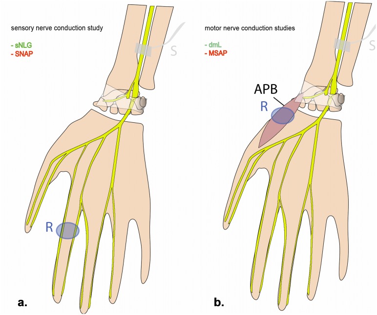
Purpose: To investigate the potential of diffusion tensor imaging (DTI) parameters as in-vivo biomarkers of axon and myelin sheath integrity of the median nerve in the carpal tunnel as validated by correlation with electrophysiology.

Methods: MRI examinations at 3T including DTI were conducted on wrists in 30 healthy subjects. After manual segmentation of the median nerve quantitative analysis of fractional anisotropy (FA) as well as axial, radial and mean diffusivity (AD, RD, and MD) was carried out. Pairwise Pearson correlations with electrophysiological parameters comprising sensory nerve action potential (SNAP) and compound muscle action potential (CMAP) as markers of axon integrity, and distal motor latency (dml) and sensory nerve conduction velocity (sNCV) as markers of myelin sheath integrity were computed. The significance criterion was set at P=0.05, Bonferroni corrected for multiple comparisons.

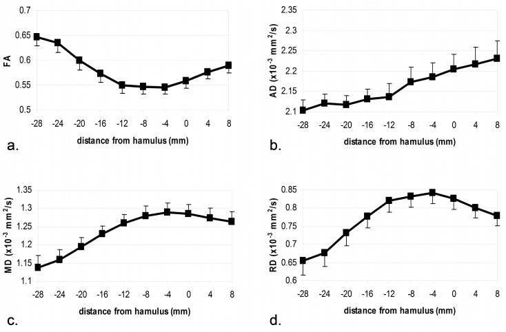
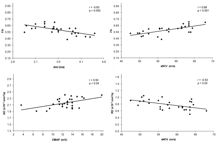
Results: DTI parameters showed a distinct proximal-to-distal profile with FA, MD, and RD extrema coinciding in the center of the carpal tunnel. AD correlated with CMAP (r=0.50, p=0.04, Bonf. corr.) but not with markers of myelin sheath integrity. RD correlated with sNCV (r=-0.53, p=0.02, Bonf. corr.) but not with markers of axon integrity. FA correlated with dml (r=-0.63, p=0.002, Bonf. corr.) and sNCV (r=0.68, p=0.001, Bonf. corr.) but not with markers of axon integrity.

Conclusion: AD reflects axon integrity, while RD (and FA) reflect myelin sheath integrity as validated by correlation with electrophysiology. DTI parameters consistently indicate a slight decrease of structural integrity in the carpal tunnel as a physiological site of median nerve entrapment. DTI is particularly sensitive, since these findings are observed in healthy participants. Our results encourage future studies to evaluate the potential of DTI in differentiating axon from myelin sheath injury in patients with manifest peripheral neuropathies.


Conflict of interest statement

Competing Interests: The authors have declared that no competing interests exist.

Figures

Fig 1
Fig 1. Schematic drawing of median nerve conduction measurements.
Sensory nerve conduction study (a): stimulation is administered at the distal forearm; sensory nerve conduction velocity (sNCV) and sensory nerve action potential (SNAP) are recorded from the digital nerve of the index finger in antidromic technique. Motor nerve conduction study (b): stimulation is administered at the distal forearm; distal motor latency (dml) and compound muscle action potential (CMAP) are recorded from the APB. Electroneurographic parameters probing axon and myelin sheath integrity are printed in red and green, respectively. APB: abductor pollicis brevis muscle; S: stimulation site; R: recording site.
Fig 2
Fig 2. DTI parameter values of the median nerve at the wrist from proximal to distal.
X-axis denotes the distance from the reference structure (hamulus of the hamate bone) in millimeters. Negative/positive values indicate position proximal/distal to that reference, respectively. Errorbars denote the standard error of the mean. FA: fractional anisotropy; AD: axial diffusivity; MD: mean diffusivity; RD: radial diffusivity.
Fig 3
Fig 3. Scatter plots relating electroneurographic parameters with DTI parameters.
Significant correlations surviving Bonferroni correction for multiple comparisons (see Table 2) are displayed. FA: fractional anisotropy; AD: axial diffusivity; RD: radial diffusivity; CMAP compound muscle action potential; dml: distal motor latency; sNCV: sensory nerve conduction velocity.

References

    1. Bendszus M, Stoll G (2005) Technology insight: visualizing peripheral nerve injury using MRI. Nat Clin Pract Neurol 1: 45–53. - PubMed
    1. Baumer P, Dombert T, Staub F, Kaestel T, Bartsch AJ, Heiland S, et al. (2011) Ulnar Neuropathy at the Elbow: MR Neurography—Nerve T2 Signal Increase and Caliber. Radiology 260: 199–206. 10.1148/radiol.11102357 - DOI - PubMed
    1. Pham M, Baumer P, Meinck HM, Schiefer J, Weiler M, Bendszus M, et al. (2014) Anterior interosseous nerve syndrome: Fascicular motor lesions of median nerve trunk. Neurology 82: 598–606. 10.1212/WNL.0000000000000128 - DOI - PMC - PubMed
    1. Takagi T, Nakamura M, Yamada M, Hikishima K, Momoshima S, Fujiyoshi K, et al. (2009) Visualization of peripheral nerve degeneration and regeneration: monitoring with diffusion tensor tractography. Neuroimage 44: 884–892. 10.1016/j.neuroimage.2008.09.022 - DOI - PubMed
    1. Lehmann HC, Zhang J, Mori S, Sheikh KA (2010) Diffusion tensor imaging to assess axonal regeneration in peripheral nerves. Exp Neurol 223: 238–244. 10.1016/j.expneurol.2009.10.012 - DOI - PMC - PubMed

Publication types