. 2015 Oct;13(10):1367-76.
doi: 10.1158/1541-7786.MCR-14-0494. Epub 2015 Jun 26.

Integration of Downstream Signals of Insulin-like Growth Factor-1 Receptor by Endoplasmic Reticulum Stress for Estrogen-Induced Growth or Apoptosis in Breast Cancer Cells

Affiliations

Integration of Downstream Signals of Insulin-like Growth Factor-1 Receptor by Endoplasmic Reticulum Stress for Estrogen-Induced Growth or Apoptosis in Breast Cancer Cells

Ping Fan et al. Mol Cancer Res. 2015 Oct.

Abstract

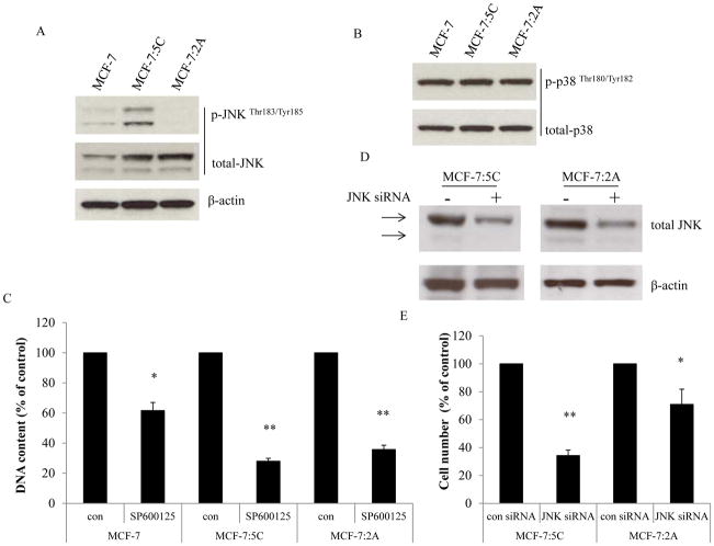
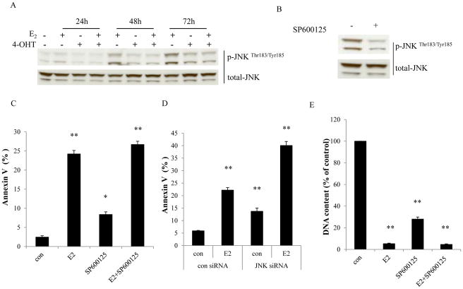
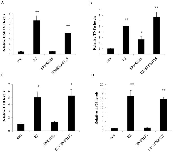
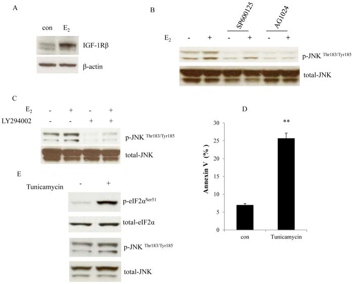
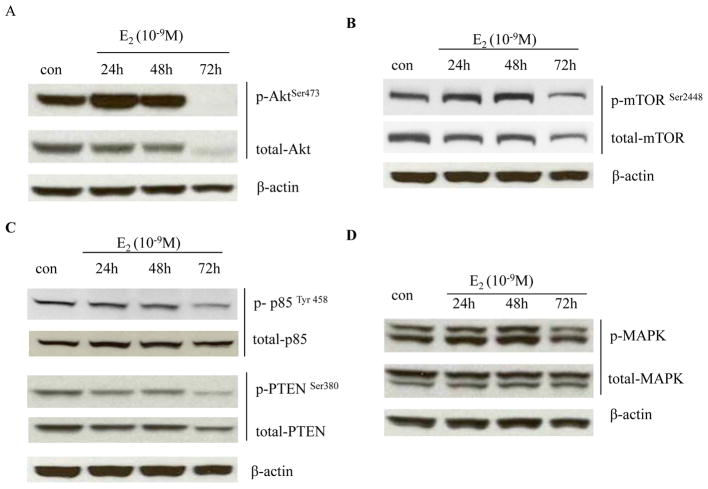
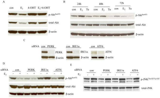
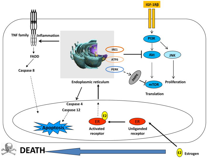
Estrogen (E2) exerts a dual function on E2-deprived breast cancer cells, with both initial proliferation and subsequent induction of stress responses to cause apoptosis. However, the mechanism by which E2 integrally regulates cell growth or apoptosis-associated pathways remains to be elucidated. Here, E2 deprivation results in many alterations in stress-responsive pathways. For instance, E2-deprived breast cancer cells had higher basal levels of stress-activated protein kinase, c-Jun N-terminal kinase (JNK), compared with wild-type MCF-7 cells. E2 treatment further constitutively activated JNK after 24 hours. However, inhibition of JNK (SP600125) was unable to abolish E2- induced apoptosis, whereas SP600125 alone arrested cells at the G2 phase of the cell cycle and increased apoptosis. Further examination showed that inhibition of JNK increased gene expression of TNFα and did not effectively attenuate expression of apoptosis-related genes induced by E2. A notable finding was that E2 regulated both JNK and Akt as the downstream signals of insulin-like growth factor-1 receptor (IGFIR)/PI3K, but with distinctive modulation patterns: JNK was constitutively activated, whereas Akt and Akt-associated proteins, such as PTEN and mTOR, were selectively degraded. Endoplasmic reticulum-associated degradation (ERAD) was involved in the selective protein degradation. These findings highlight a novel IGFIR/PI3K/JNK axis that plays a proliferative role during the prelude to E2-induced apoptosis and that the endoplasmic reticulum is a key regulatory site to decide cell fate after E2 treatment.

Implications: This study provides a new rationale for further exploration of E2-induced apoptosis to improve clinical benefit.


Conflict of interest statement

There are no conflicts to disclose.

Figures

Figure 1
Figure 1. Basal levels of JNK and p38 in three cell lines
(A) Expression levels of JNK in three cell lines. MCF-7 cells were cultured in E2-free medium for three days. Then, cell lysates of MCF-7, MCF-7:5C, and MCF-7:2A were harvested. Total JNK and p-JNK were examined by Western blotting. β-actin was measured as loading control. (B) Expression levels of p38 in three cell lines. Cell lysates of MCF-7, MCF-7:5C, and MCF-7:2A were the same as above. Total p38 and p-p38 were examined by Western blotting. (C) Growth response to the JNK inhibitor. MCF-7, MCF-7:5C, and MCF-7:2A cells were treated with vehicle (0.1% DMSO) or SP600125 (10−5 mol/L). Cells were harvested after 7 days of treatment and cell viability was quantitated by determination of total DNA. p<0.05, * compared with control. p<0.001, ** compared with control. (D) Knockdown of JNK through specific siRNA. MCF-7:5C cells were transfected with control siRNA or JNK siRNA for 72 hours. MCF-7:2A cells were double transfected with control siRNA or JNK siRNA for 5 days. Cell lysates were harvested for Western blotting. (E) Knockdown of JNK inhibited cell growth. MCF-7:5C and MCF-7:2A cells were double transfected with control siRNA or JNK siRNA and were grown for 5 days. Cell nuclei were counted using a Coulter counter. p<0.05, * compared with control. p<0.001, ** compared with control.
Figure 2
Figure 2. Activation of JNK by E2 in MCF-7:5C cells
(A) Activation of JNK by E2. MCF-7:5C cells were treated with vehicle (0.1% EtOH), E2 (10−9 mol/L), 4-OHT (10−6 mol/L), and E2 (10−9 mol/L) plus 4-OHT (10−6 mol/L) for the time points indicated. p-JNK was examined by Western blotting. Total JNK was measured as loading control. (B) The JNK inhibitor effectively blocked phosphorylation of JNK. MCF-7:5C cells were treated with vehicle (0.1% DMSO) or SP600125 (10−5 mol/L) for 24 hours. p-JNK was examined by Western blotting. Total JNK was measured as loading control. (C) The JNK inhibitor could not block E2-induced apoptosis. MCF-7:5C cells were treated with vehicle (0.1% DMSO), E2 (10−9 mol/L), SP600125 (10−5 mol/L), and E2 (10−9 mol/L) plus SP600125 (10−5 mol/L) for 72 hours. Annexin V binding assay was used to detect apoptosis. p<0.05, * compared with control. p<0.001, ** compared with control. (D) Knockdown of JNK could not block E2-induced apoptosis. MCF-7:5C cells were transfected with control siRNA or JNK siRNA for 72 hours. Then, cells were treated with vehicle (0.1% EtOH) or E2 (10−9 mol/L) for 72 hours. Annexin V binding assay was used to detect apoptosis. p<0.001, ** compared with control. (E) Growth response to the JNK inhibitor in the presence or absence of E2. MCF-7:5C cells were treated with the same compound as in (C). Cells were harvested after 7 days treatment and cell viability was quantitated by determination of total DNA. p<0.001, ** compared with control.
Figure 3
Figure 3. Regulation of apoptosis-related genes by the JNK inhibitor
MCF-7:5C cells were treated with the same compounds as in Fig. 2C for 72 hours. Cells were harvested in TRIzol. Gene expression levels were quantitated by RT-PCR. (A) HMOX1, (B) TNF, (C) LTB, (D) TP63. p<0.05, * compared with control. p<0.001, ** compared with control.
Figure 4
Figure 4. Regulation of the JNK activation
(A) E2 elevated IGF-1R. MCF-7:5C cells were treated with vehicle (0.1% EtOH) or E2 (10−9 mol/L) for 72 hours. IGF-1R was examined by Western blotting. β-actin was measured as loading control. (B) Regulation of JNK activation by IGF-1R. MCF-7:5C cells were treated with vehicle (0.1% DMSO), E2 (10−9 mol/L), SP600125 (10−5 mol/L), E2 (10−9 mol/L) plus SP600125 (10−5 mol/L), AG1024 (5×10⁻⁶ mol/L), E2 (10−9 mol/L) plus AG1024 (5×10⁻⁶ mol/L) for 48 hours. p-JNK was examined by Western blotting. Total JNK was measured as loading control. (C) Regulation of JNK activation by PI3K. MCF-7:5C cells were treated with vehicle (0.1% DMSO), E2 (10−9 mol/L), LY294002 (10−6 mol/L), and E2 (10−9 mol/L) plus LY294002 (10−6 mol/L) for 48 hours. p-JNK was examined by Western blotting. Total JNK was measured as loading control. (D) Induction of apoptosis by the endoplasmic reticulum stress inducer, Tunicamycin. MCF-7:5C cells were treated with vehicle (0.1% DMSO) or Tunicamycin (10−5 mol/L) for 24 hours. Annexin V binding assay was used to detect apoptosis. p<0.001, ** compared with control. (E) Effects of Tunicamycin on the activation of JNK. MCF-7:5C cells were treated with the same compound as in (D). p-eIF2α and p-JNK were examined by Western blotting. Total eIF2α and JNK were measured as loading controls.
Figure 5
Figure 5. E2 degraded Akt-associated proteins
(A) Regulation of Akt by E2. MCF-7:5C cells were treated with vehicle (0.1% EtOH) or E2 (10−9 mol/L) for different time points indicated. Cell lysates were harvested. p-Akt and total Akt were examined by Western blotting. β-actin was measured as loading control. (B) Regulation of mTOR by E2. Cells were treated the same as in (A). p-mTOR and total mTOR were examined by Western blotting. β-actin was measured as loading control. (C) Regulation of PTEN and p85 by E2. Cell lysates were the same as above. p-PTEN, total PTEN, p-p85, and total p85 were examined by Western blotting. β-actin was measured as loading control. (D) Regulation of MAPK by E2. MCF-7:5C cells were treated with vehicle (0.1% EtOH) or E2 (10−9 mol/L) for different time points indicated. Cell lysates were harvested. p-MAPK and total MAPK were examined by Western blotting. β-actin was measured as loading control.
Figure 6
Figure 6. Regulation of Akt by endoplasmic reticulum stress
(A) 4-OHT prevented the degradation of Akt by E2. MCF-7:5C cells were treated with vehicle (0.1% EtOH), E2 (10−9 mol/L), 4-OHT (10−6 mol/L), and 4-OHT (10−6 mol/L) plus E2 (10−9 mol/L) for 72 hours. Cell lysates were harvested. p-Akt and total Akt were examined by Western blotting. β-actin was measured as loading control. (B) Effects of Tunicamycin on Akt phosphorylation. MCF-7:5C cells were treated with vehicle (0.1% DMSO), E2 (10−9 mol/L), and Tunicamycin (10−5 mol/L) for different time points indicated. p-Akt and total-Akt were examined by Western blotting. β-actin was measured as loading control. (C) Knockdown of three sensors of endoplasmic reticulum stress by siRNAs. MCF-7:5C cells were transfected with scrambled, PERK, IRE1α, and ATF6 siRNA for 72 hours. Respective proteins were examined by Western blotting. β-actin was measured as loading control. (D) Effects on Akt after knockdown of three sensors. MCF-7:5C cells were transfected with specific siRNAs as above for 72 hours. Then, cells were treated with or without E2 for 72 hours. p-Akt and total-Akt were examined by Western blotting. β-actin was measured as loading control. (E) Effects on JNK phosphorylation after knockdown of three sensors. Cell lysates were the same as in (D). p-JNK and total-JNK were examined by Western blotting.
Figure 7
Figure 7. Endoplasmic reticulum is a joint regulatory site to integrally modulate growth or apoptosis associated pathways
E2 activates IGF-1R/PI3K and its downstream signals, Akt and JNK to promote cell growth. Simultaneously, E2 activates endoplasmic reticulum stress which activates a set of signaling pathways including three sensors (PERK, IRE1α, and ATF6), inflammatory responses, caspase 4/12, and adenosine monophosphate (AMP)-activated protein kinase (AMPK). The AMPK and Akt converge on mTOR with opposing regulatory effects to coordinate bioenergetics and cell viability. IRE1α and ATF6 are involved in the degradation of Akt.

References

    1. Jordan VC, Brodie AM. Development and evolution of therapies targeted to the estrogen receptor for the treatment and prevention of breast cancer. Steroids. 2007;72:7–25. - PMC - PubMed
    1. Wolf DM, Jordan VC. A laboratory model to explain the survival advantage observed in patients taking adjuvant tamoxifen therapy. Recent Results Cancer Res. 1993;127:23–33. - PubMed
    1. Yao K, Lee ES, Bentrem DJ, England G, Schafer JI, O’Regan RM, et al. Antitumor action of physiological estradiol on tamoxifen-stimulated breast tumors grown in athymic mice. Clin Cancer Res. 2000;6:2028–36. - PubMed
    1. Song RX, Mor G, Naftolin F, McPherson RA, Song J, Zhang Z, et al. Effect of long-term estrogen deprivation on apoptotic responses of breast cancer cells to 17beta-estradiol. J Natl Cancer Inst. 2001;93:1714–23. - PubMed
    1. Lewis JS, Meeke K, Osipo C, Ross EA, Kidawi N, Li T, et al. Intrinsic mechanism of estradiol-induced apoptosis in breast cancer cells resistant to estrogen deprivation. J Natl Cancer Inst. 2005;97:1746–59. - PubMed

Publication types

MeSH terms