. 2015 Aug 15;128(16):2983-8.
doi: 10.1242/jcs.168179. Epub 2015 Jun 26.

Endothelial MMP14 is required for endothelial-dependent growth support of human airway basal cells

Affiliations

Endothelial MMP14 is required for endothelial-dependent growth support of human airway basal cells

Bi-Sen Ding et al. J Cell Sci. .

Abstract

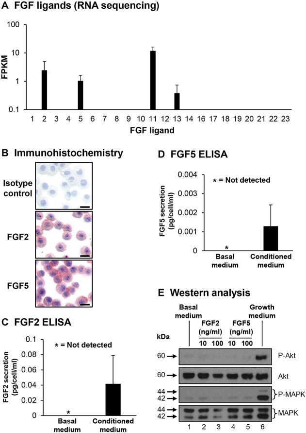
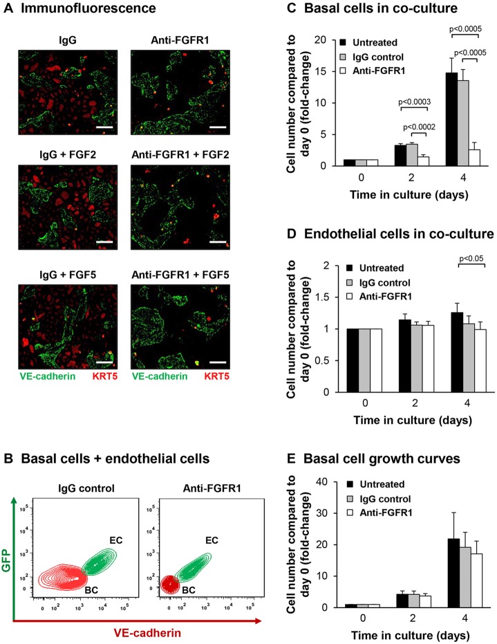
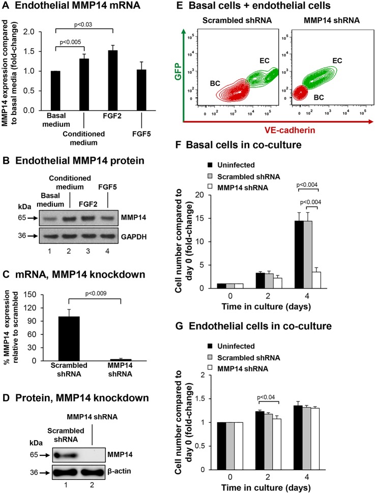
Human airway basal cells are the stem (or progenitor) population of the airway epithelium, and play a central role in anchoring the epithelium to the basement membrane. The anatomic position of basal cells allows for potential paracrine signaling between them and the underlying non-epithelial stromal cells. In support of this, we have previously demonstrated that endothelial cells support growth of basal cells during co-culture through vascular endothelial growth factor A (VEGFA)-mediated signaling. Building on these findings, we found, by RNA sequencing analysis, that basal cells expressed multiple fibroblast growth factor (FGF) ligands (FGF2, FGF5, FGF11 and FGF13) and that only FGF2 and FGF5 were capable of functioning in a paracrine manner to activate classical FGF receptor (FGFR) signaling. Antibody-mediated blocking of FGFR1 during basal-cell-endothelial-cell co-culture significantly reduced the endothelial-cell-dependent basal cell growth. Stimulation of endothelial cells with basal-cell-derived growth factors induced endothelial cell expression of matrix metallopeptidase 14 (MMP14), and short hairpin RNA (shRNA)-mediated knockdown of endothelial cell MMP14 significantly reduced the endothelial-cell-dependent growth of basal cells. Overall, these data characterize a new growth-factor-mediated reciprocal 'crosstalk' between human airway basal cells and endothelial cells that regulates proliferation of basal cells.

Keywords: Airway basal cell; Crosstalk; Endothelial cell; MMP14; Progenitor cell.


Conflict of interest statement

Competing interests

The authors declare that they have no competing interests, but notify that S.R. is the founder of and consultant to Angiocrine Bioscience New York, NY, USA.

Figures

Fig. 1.
Fig. 1.
Basal cell expression of FGF family ligands. (A) RNA sequencing analysis of FGF ligands in basal cells. Data shown represents the mean±s.d FPKM expression from n=10 independent samples. (B) Immunohistochemical staining of cytopreparations of basal cells for FGF2, FGF5 and an isotype control. Scale bar: 20 µm. (C,D) FGF2 and FGF5 levels assessed by ELISA in medium from basal cells. Secreted FGF2 and FGF5 were normalized to the cell number and calculated as pg per cell/ml. Data shown is the mean±s.d of three independent samples, each performed in triplicate. (E) Airway basal cells were starved of growth factors for 6 h and then stimulated for 15 min with basal medium (lane 1), basal medium containing FGF2 (10 and 100 ng/ml) (lanes 2 and 3) or FGF5 (10 and 100 ng/ml) (lanes 4 and 5), or growth-factor-containing medium (lane 6). Activation of Akt and MAPK signaling was evaluated by western blot analysis and staining for phosphorylated Akt (P-Akt) and phosphorylated ERK1/2 (P-MAPK). The levels of total Akt and ERK1/2 (MAPK) were evaluated as a loading control.
Fig. 2.
Fig. 2.
Blocking of FGFR1 suppresses endothelial cells-dependent proliferation of basal cells. (A–D) Airway basal cells were co-cultured with endothelial cells (HUVEC-Akt) and incubated with control IgG or anti-FGFR1 antibody. For A, FGF2 (10 ng/ml) or FGF5 (10 ng/ml) were also added. (A) Immunofluorescence of basal cells and endothelial cells in co-culture. Basal cells were identified by KRT5 staining (red) and endothelial cells were identified by VE-cadherin staining (green). Scale bars: 100 µm. (B) Representative flow cytometric analysis of basal cells (BC) and endothelial cells (EC) in co-culture. (C) Proliferation of basal cells co-cultured with endothelial cells. (D) Proliferation of endothelial cells in co-culture with basal cells. (E) Proliferation of basal cells cultured alone in growth medium and incubated with control IgG or anti-FGFR1 antibody. For C–E, shown are untreated (black), IgG (gray), and anti-FGFR1 antibody (white) data; results are the mean±s.d. of four independent experiments, each performed in triplicate.
Fig. 3.
Fig. 3.
Endothelial cell MMP14 is required for endothelial-cell-dependent induction of basal cell proliferation. (A,B) Basal-cell-derived growth factors stimulate expression of endothelial cells MMP14. (A) mRNA (TaqMan) analysis of MMP14 expression in growth-factor-starved endothelial cells (HUVECs) stimulated with either basal medium, basal medium conditioned on basal cells, or basal medium containing FGF2 (10 ng/ml) or FGF5 (10 ng/ml). Data shown is the mean±s.d. of at least three independent experiments, each performed in triplicate. (B) Protein levels of MMP14 in cells treated as in A. A representative western blot analysis is shown. (C,D) shRNA-mediated knockdown of endothelial cell (HUVEC-Akt) MMP14 expression. (C) mRNA analysis (TaqMan) of MMP14 expression in cells infected with the indicated shRNA. (D) Protein levels of MMP14 in cells treated as in C. A representative western blot analysis is shown. (E–G) Analysis of basal cell and endothelial cell co-culture. (E) Representative flow cytometric analysis of basal cells (BC) and endothelial cells (EC) infected with scrambled or MMP14 shRNA cells in co-culture. (F) Basal cell number in co-culture. (G) Endothelial cell numbers in co-culture. For F–G, shown are the endothelial cells (black), endothelial cells containing scrambled shRNA (gray) and MMP14 shRNA (white) data; results are the mean±s.d. of three independent experiments each performed in triplicate.

References

    1. Allerstorfer S., Sonvilla G., Fischer H., Spiegl-Kreinecker S., Gauglhofer C., Setinek U., Czech T., Marosi C., Buchroithner J., Pichler J. et al. (2008). FGF5 as an oncogenic factor in human glioblastoma multiforme: autocrine and paracrine activities. Oncogene 27, 4180-4190. 10.1038/onc.2008.61 - DOI - PMC - PubMed
    1. Belov A. A. and Mohammadi M. (2013). Molecular mechanisms of fibroblast growth factor signaling in physiology and pathology. Cold Spring Harb. Perspect. Biol. 5, a015958 10.1101/cshperspect.a015958 - DOI - PMC - PubMed
    1. Carter E. P., Fearon A. E. and Grose R. P. (2015). Careless talk costs lives: fibroblast growth factor receptor signalling and the consequences of pathway malfunction. Trends Cell Biol. 25, 221-233. 10.1016/j.tcb.2014.11.003 - DOI - PubMed
    1. Crystal R. G., Randell S. H., Engelhardt J. F., Voynow J. and Sunday M. E. (2008). Airway epithelial cells: current concepts and challenges. Proc. Am. Thorac. Soc. 5, 772-777. 10.1513/pats.200805-041HR - DOI - PMC - PubMed
    1. Curradi G., Walters M. S., Ding B.-S., Rafii S., Hackett N. R. and Crystal R. G. (2012). Airway basal cell vascular endothelial growth factor-mediated cross-talk regulates endothelial cell-dependent growth support of human airway basal cells. Cell. Mol. Life Sci. 69, 2217-2231. 10.1007/s00018-012-0922-8 - DOI - PMC - PubMed

Publication types

MeSH terms