. 2015 Sep 1;81(17):6078-87.
doi: 10.1128/AEM.01254-15. Epub 2015 Jun 26.

Type III Secretion System Translocon Component EseB Forms Filaments on and Mediates Autoaggregation of and Biofilm Formation by Edwardsiella tarda

Affiliations

Type III Secretion System Translocon Component EseB Forms Filaments on and Mediates Autoaggregation of and Biofilm Formation by Edwardsiella tarda

Zhi Peng Gao et al. Appl Environ Microbiol. .

Abstract

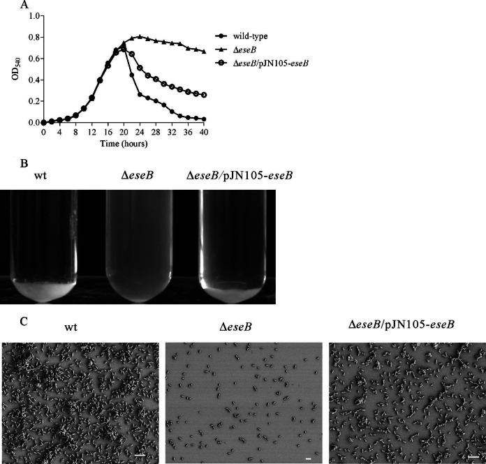
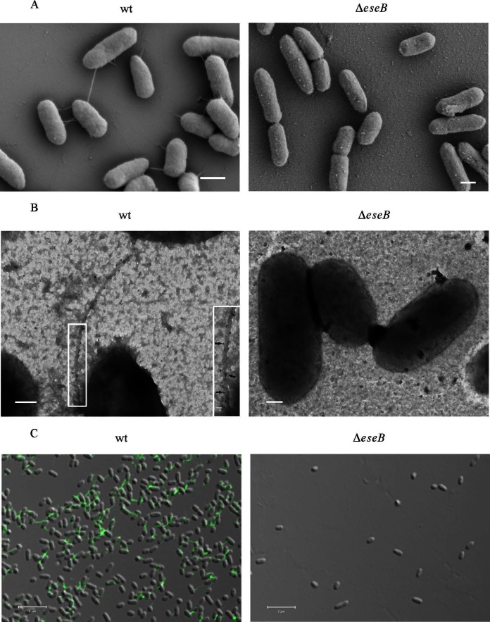
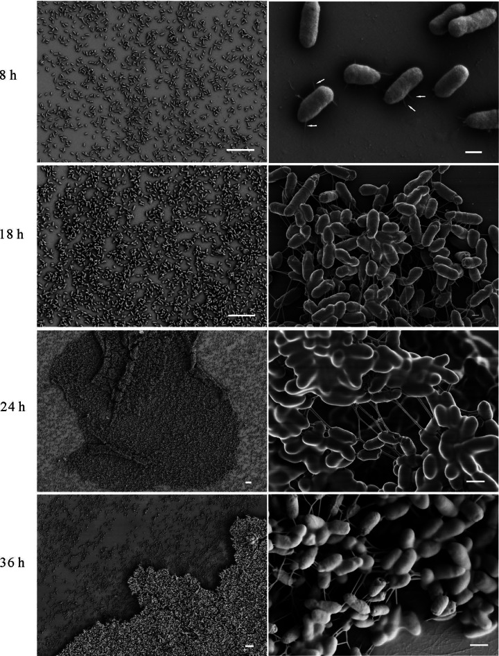
The type III secretion system (T3SS) of Edwardsiella tarda plays an important role in infection by translocating effector proteins into host cells. EseB, a component required for effector translocation, is reported to mediate autoaggregation of E. tarda. In this study, we demonstrate that EseB forms filamentous appendages on the surface of E. tarda and is required for biofilm formation by E. tarda in Dulbecco's modified Eagle's medium (DMEM). Biofilm formation by E. tarda in DMEM does not require FlhB, an essential component for assembling flagella. Dynamic analysis of EseB filament formation, autoaggregation, and biofilm formation shows that the formation of EseB filaments occurs prior to autoaggregation and biofilm formation. The addition of an EseB antibody to E. tarda cultures before bacterial autoaggregation prevents autoaggregation and biofilm formation in a dose-dependent manner, whereas the addition of the EseB antibody to E. tarda cultures in which biofilm is already formed does not destroy the biofilm. Therefore, EseB filament-mediated bacterial cell-cell interaction is a prerequisite for autoaggregation and biofilm formation.


Figures

FIG 1
FIG 1
Dynamic analysis of E. tarda autoaggregation. (A) EseB-mediated E. tarda autoaggregation. Deletion of eseB abolished E. tarda autoaggregation, and expression of eseB in pJN105-eseB induced by 50 mM l-arabinose restored autoaggregation to wild-type levels. (B) Autoaggregation of wild-type (wt), ΔeseB, and ΔeseB/pJN105-eseB cells in DMEM at 25°C. The wild-type and the complementing strains began to autoaggregate at 20 hps. (C) Autoaggregation observed by SEM. Cell clumps that settled on coverslips were observed for the wild-type and ΔeseB/pJN105-eseB strains but not the ΔeseB strain. Bars, 4 μm.
FIG 2
FIG 2
EseB forms filamentous appendages on the surface of E. tarda cells. (A) SEM of E. tarda wild-type strain PPD130/91 and the ΔeseB strain. Bars, 600 nm. (B) Immuno-TEM images of the E. tarda wild-type and ΔeseB strains. Bacteria were labeled with anti-EseB antibody and protein A-coated colloidal gold particles conjugated to donkey anti-mouse secondary antibody (10 nm in diameter). Gold particles were distributed along filamentous appendages on E. tarda wild-type (black arrows) but not on ΔeseB cells. The inset shows enlarged views of the boxed areas. Bars, 200 nm; bar for the inset, 20 nm. (C) Immunofluorescence staining of wild-type and ΔeseB cells with antibody against EseB. The fixed bacteria were incubated with anti-EseB antibody, followed by incubation with Alexa 488 donkey anti-mouse secondary antibody. Green filamentous signals were detected in wild-type bacteria but not in ΔeseB strain bacteria. Bars, 5 μm.
FIG 3
FIG 3
EseB but not FlhB is involved in E. tarda biofilm formation in DMEM. (A) Deletion of flhB does not influence E. tarda biofilm formation in DMEM. Biofilms that developed were stained with crystal violet, and biofilm formation was evaluated by examining the OD630 of the dissolved crystal violet. *** indicates a significant difference at a P value of <0.001. (B) Motility of wild-type, ΔeseB, and ΔflhB cells on TSA with 0.4% agar. Images of motility halos were taken 12 h after loading of bacteria.
FIG 4
FIG 4
Dynamic analysis of biofilm formation and EseB filaments. At 8 h, 18 h, 24 h, and 36 h postsubculture, E. tarda wild-type cells that settled on the coverslips were assayed by SEM. At 8 hps, EseB helps E. tarda to attach to the coverslips, as indicated by the white arrows, and at 24 and 36 hps, EseB helps to connect and support E. tarda cells. Bars, 10 μm at low magnification (left) and 400 nm at high magnification (right).
FIG 5
FIG 5
EseB antibody blocks E. tarda autoaggregation and biofilm formation. E. tarda wild-type cells cultured in DMEM in tubes or 24-well plates were supplemented (Treated) with EseB antibody at 1:2,000, 1:1,000, 1:200, 1:100, and 1:50 dilutions. Naive mouse serum was added to E. tarda wild-type cultures at a 1:50 dilution and is labeled Untreated. (A) EseB antibody added at 15 hps inhibits autoaggregation of E. tarda cultured in DMEM in a dose-dependent manner, as shown by OD540 values of the culture supernatants and images of autoaggregation in glass tubes. *** , P < 0.001; **, P < 0.01. (B) Biofilm formation by E. tarda cultured in DMEM is inversely proportional to the amount of EseB antibody added at 15 hps. Crystal violet staining and quantification of dissolved crystal violet were performed to evaluate the biofilm formation ability in the presence of EseB antibody. ***, P < 0.001. (C) Confocal laser scanning microscopy images of top-down views and orthogonal views of filament formation when EseB antibody was added at 15 hps. There were fewer EseB filaments with increasing concentrations of EseB antibody. The amounts of EseB antibody added are indicated for dilutions of no less than 1:100. (D) Mature biofilm formed by E. tarda is not influenced by EseB antibody added at 24 hps. The biofilms were examined by crystal violet staining at 4 h post-EseB antibody supplementation. The quality and amount of biofilm formation were revealed by crystal violet staining. ***, P < 0.001.

References

    1. Manchanda V, Singh NP, Eideh HK, Shamweel A, Thukral SS. 2006. Liver abscess caused by Edwardsiella tarda biogroup 1 and identification of its epidemiological triad by ribotyping. Indian J Med Microbiol 24:135–137. doi:10.4103/0255-0857.25205. - DOI - PubMed
    1. Leung KY, Siame BA, Tenkink BJ, Noort RJ, Mok Y-K. 2012. Edwardsiella tarda—virulence mechanisms of an emerging gastroenteritis pathogen. Microbes Infect 14:26–34. doi:10.1016/j.micinf.2011.08.005. - DOI - PubMed
    1. Koido S, Ohkusa T, Kato K, Shimamoto N, Takakura K, Odahara S, Tsukinaga S, Mitobe J, Yukawa T, Kajihara M, Uchiyama K, Arakawa H, Tajiri H. 2014. Edwardsiella tarda superinfection in relapse of ulcerative colitis. J Clin Gastroenterol 48:191–193. doi:10.1097/01.mcg.0000437809.46982.df. - DOI - PubMed
    1. Janda JM, Abbott SL, Oshiro L. 1991. Penetration and replication of Edwardsiella spp. in HEp-2 cells. Infect Immun 59:154–161. - PMC - PubMed
    1. Tan YP, Zheng J, Tung SL, Rosenshine I, Leung KY. 2005. Role of type III secretion in Edwardsiella tarda virulence. Microbiology 151:2301–2313. doi:10.1099/mic.0.28005-0. - DOI - PubMed

Publication types

MeSH terms

Substances

LinkOut - more resources