. 2015 Sep 1;405(1):1-9.
doi: 10.1016/j.ydbio.2015.06.005. Epub 2015 Jun 24.

A thioredoxin fold protein Sh3bgr regulates Enah and is necessary for proper sarcomere formation

Affiliations

A thioredoxin fold protein Sh3bgr regulates Enah and is necessary for proper sarcomere formation

Dong Gil Jang et al. Dev Biol. .

Abstract

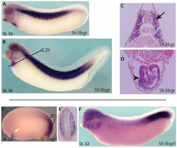
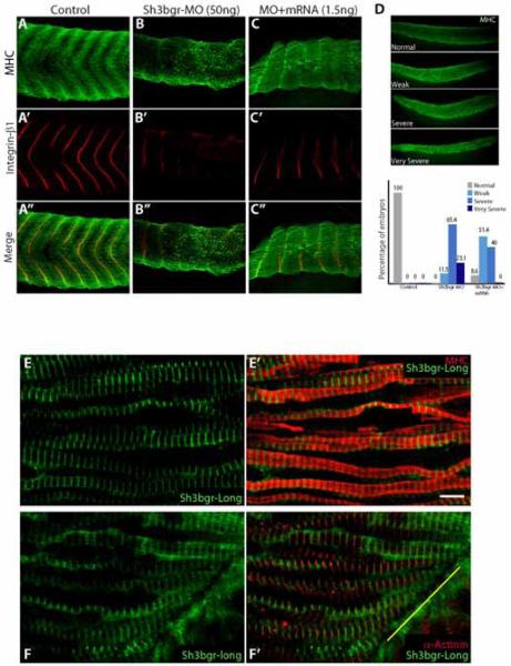
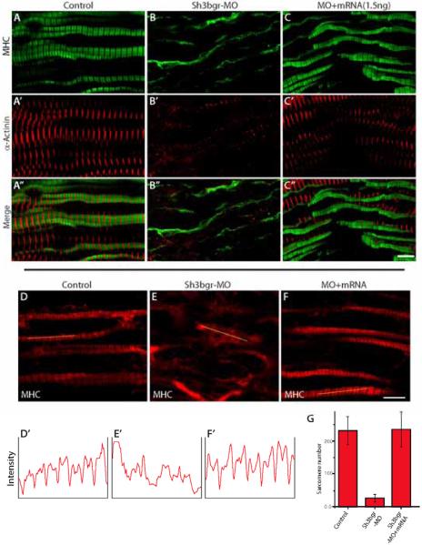
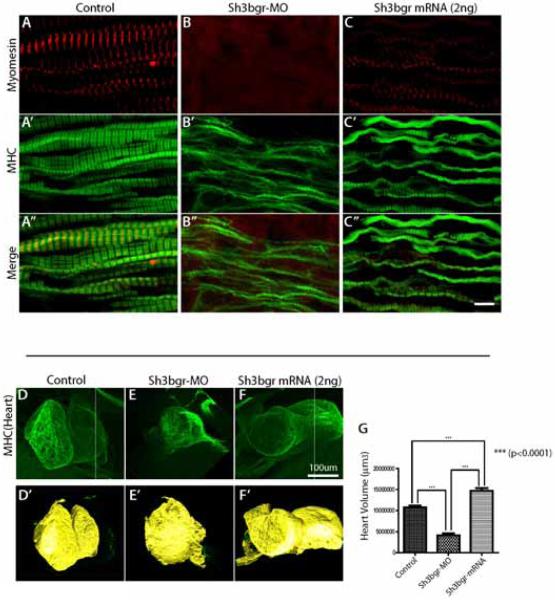
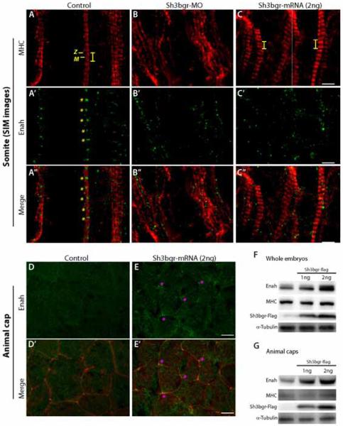
The sh3bgr (SH3 domain binding glutamate-rich) gene encodes a small protein containing a thioredoxin-like fold, SH3 binding domain, and glutamate-rich domain. Originally, it was suggested that increased expression of Sh3bgr may cause the cardiac phenotypes in Down's syndrome. However, it was recently reported that the overexpression of Sh3bgr did not cause any disease phenotypes in mice. In this study, we have discovered that Sh3bgr is critical for sarcomere formation in striated muscle tissues and also for heart development. Sh3bgr is strongly expressed in the developing somites and heart in Xenopus. Morpholino mediated-knockdown of sh3bgr caused severe malformation of heart tissue and disrupted segmentation of the somites. Further analysis revealed that Sh3bgr specifically localized to the Z-line in mature sarcomeres and that knockdown of Sh3bgr completely disrupted sarcomere formation in the somites. Moreover, overexpression of Sh3bgr resulted in abnormally discontinues thick firmaments in the somitic sarcomeres. We suggest that Sh3bgr does its function at least partly by regulating localization of Enah for the sarcomere formation. In addition, we provide the data supporting Sh3bgr is also necessary for proper heart development in part by affecting the Enah protein level.


Figures

Figure 1
Figure 1. sh3bgr is expressed in the skeletal muscles and heart tissues
Whole mount in situ hybridization showing that sh3bgr is expressed in the somites and heart at Stage 30 (A), Stage 36 (B). C, D. Sectioned samples shown in (B). E. sh3bgrl is expressed in the presomitic tissue and early cardiac tissue at Stage 23. F. At Stage 32, sh3bgrl is expressed in the pharyngeal arches and the presomitic expression was maintained.
Figure 2
Figure 2. Knockdown of sh3bgr caused severe defects in somite development
A-C. Microinjection of sh3bgr antisense morpholino (Sh3bgr-MO; 50ng per embryo) disrupted normal somite development. The somite boundaries were labeled with anti-integrin-β1 antibody (Red) and the muscle fibers were stained with the myosin heavy chain (MHC) antibody (Green) at Stage 33. D. The muscle phenotypes were categorized according to the severity. Weak; the chevron pattern is partially disrupted, Severe; the chevron pattern is completely lost. Very severe; the somatic tissue is severely hypomorphic. The muscle defects in the morphants were recovered by co-injection with 1.5ng of sh3bgr mRNA. The numbers in the graph are the percentage of the embryos. The numbers of embryos analyzed: Control-13, sh3bgr morphants-26, Rescue-35. E-E’. Sh3bgr localizes to the muscle fibers. 150ng of Sh3bgr-flag mRNA was injected into Stage 2 embryos. Stage 33 embryos were fixed and the coronal sections of somitic tissues were stained for MHC (Red) and Sh3bgr-flag (Green). F-F’. Sh3bgr-flag fusion protein localized to the Z-line in sarcomeres. Sh3bgr-flag (Green) was co-immunostained with α-actinin (Red). The yellow line indicates a somitic boundary. Scale bar = 5μm
Figure 3
Figure 3. sh3bgr is necessary for sarcomere formation
A-C. Stage 33 embryos were fixed and the coronal sections of somitic tissues were stained for α-actinin (Red) and MHC (Green). Knockdown of sh3bgr caused severe defects in the sarcomeres. Sh3bgr knockdown caused almost compete loss of sarcomere structures (B). Co-injection of Sh3bgr mRNA (1.5ng) with Sh3bgr-MO rescued sarcomere defects (C). D-F. Representative images of thick filaments. The fluorescent intensity plots of indicated muscle fibers are shown in (D’-F’). The average numbers of intact sarcomeres in each sample are shown in (G). Scale bar = 5μm
Figure 4
Figure 4. Overexpression of Sh3bgr caused abnormal sarcomere formation and increased heart volume
A-C. Stage 33 embryos were fixed and the coronal sections of somitic tissues were stained for myomesin (Red) and MHC (Green). Overexpression of sh3bgr (2ng of Sh3bgr-flag mRNA) caused abnormally discontinuous thick filaments and mislocalizaiton of myomesin. D-F. Overexpression of Sh3bgr increased the heart volumes. The embryonic hearts were stained with anti-MHC antibody and the volumes of Stage 45 embryos were measured using the Imaris program. D’-F’. The surface views of 3D confocal images in (D-F) rendered using the Imaris program. The heart volume was plotted in (G). The numbers of embryos analyzed: Control-13, sh3bgr morphants-13, Rescue-13. Scale bar = 5μm
Figure 5
Figure 5. Sh3bgr is an upstream regulator of Enah
A-C. Super resolution SIM images of sarcomeres in control (A) Sh3bgr knockdown (B), and Sh3bgr overexpressing embryos (C). Enah protein was labeled in green and MHC was labeled in red. The Z-line and M-band are indicated within a sarcomere. Both knockdown and overexpression of Sh3bgr delocalize Enah from the Z-lines. D-E. Enah staining in control (D) and Sh3bgr overexpressing (E) animal cap cells. In Sh3bgr overexpressing animal cap cells, the Enah protein is highly enriched in tri-junctional foci (asterisk). F-G. The western blot analysis showed that the endogenous Enah protein level was sharply increased in whole embryos (F) or animal cap tissues (G) upon overexpression of Sh3bgr. Scale bar (A-C) = 2μm Scale bar (D, E) = 10μm

References

    1. Agarkova I, Perriard JC. The M-band: an elastic web that crosslinks thick filaments in the center of the sarcomere. Trends Cell Biol. 2005;15:477–485. - PubMed
    1. Aguilar F, Belmonte SL, Ram R, Noujaim SF, Dunaevsky O, Protack TL, Jalife J, Todd Massey H, Gertler FB, Blaxall BC. Mammalian enabled (Mena) is a critical regulator of cardiac function. American journal of physiology. Heart and circulatory physiology. 2011;300:H1841–1852. - PMC - PubMed
    1. Bear JE, Gertler FB. Ena/VASP: towards resolving a pointed controversy at the barbed end. J Cell Sci. 2009;122:1947–1953. - PMC - PubMed
    1. Belmonte SL, Ram R, Mickelsen DM, Gertler FB, Blaxall BC. Cardiac overexpression of Mammalian enabled (Mena) exacerbates heart failure in mice. American journal of physiology. Heart and circulatory physiology. 2013;305:H875–884. - PMC - PubMed
    1. Benz PM, Merkel CJ, Offner K, Abesser M, Ullrich M, Fischer T, Bayer B, Wagner H, Gambaryan S, Ursitti JA, Adham IM, Linke WA, Feller SM, Fleming I, Renne T, Frantz S, Unger A, Schuh K. Mena/VASP and alphaII-Spectrin complexes regulate cytoplasmic actin networks in cardiomyocytes and protect from conduction abnormalities and dilated cardiomyopathy. Cell Commun Signal. 2013;11:56. - PMC - PubMed

Publication types

MeSH terms

LinkOut - more resources