. 2015 Aug 7;290(32):19967-75.
doi: 10.1074/jbc.M115.661504. Epub 2015 Jun 27.

Oncogenic miR-17/20a Forms a Positive Feed-forward Loop with the p53 Kinase DAPK3 to Promote Tumorigenesis

Affiliations

Oncogenic miR-17/20a Forms a Positive Feed-forward Loop with the p53 Kinase DAPK3 to Promote Tumorigenesis

Zhiqiang Cai et al. J Biol Chem. .

Abstract

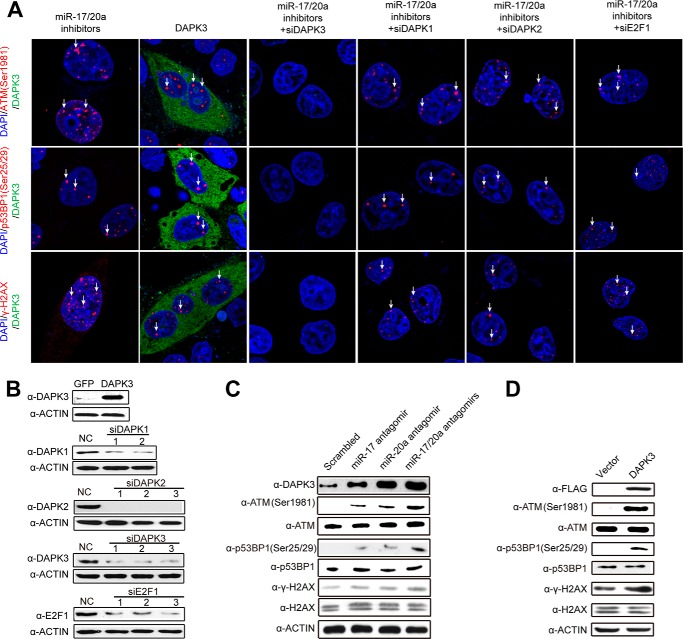
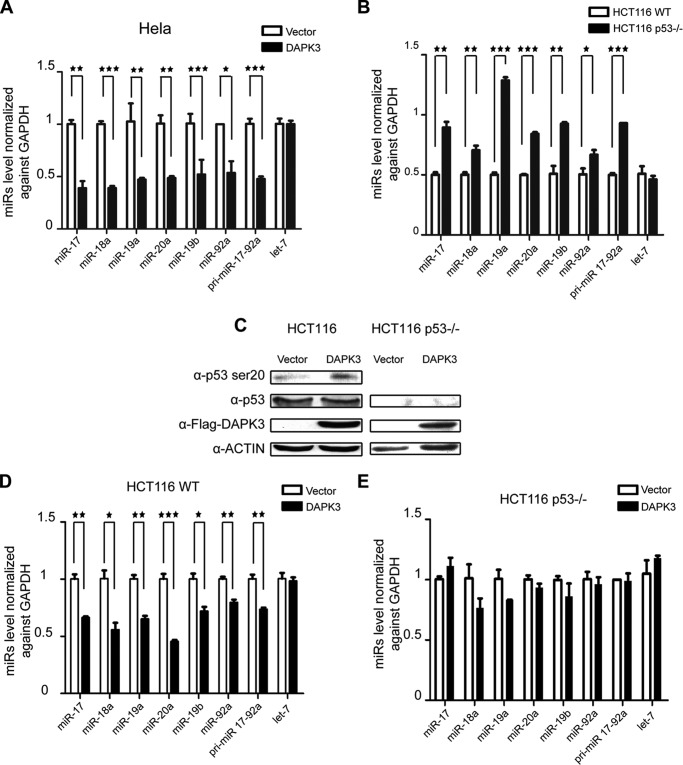
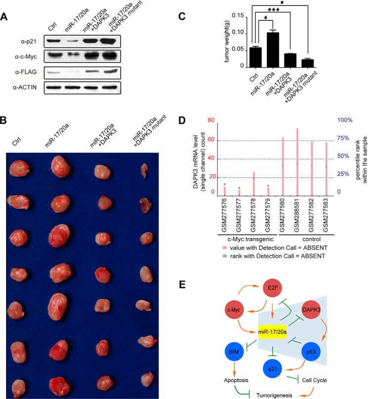
MicroRNAs (miRs) are a class of small regulatory RNAs that have been implicated in diverse biological pathways, including cancer. miR-17/20a encoded by the c13orf25 locus is among the first miRs discovered to have oncogenic functions. The E2F family members have been established as the targets for these oncomiRs, which form a negative feedback loop to control cell cycle progression. However, this pathway does not seem to be sufficient to account for elevated expression of these oncomiRs in cancer cells to promote tumorigenesis. Here we report that miR-17/20a targets a p53 activating kinase DAPK3, leading to p53-dependent transcriptional de-repression of the oncomiRs. We demonstrate that DAPK3 plays a central role in preventing miR-17/20a depletion-induced genome instability and in miR-17/20a overexpression-triggered tumor formation. This newly identified tumorigenic pathway may thus contribute to miR-17/20a amplification and tumor growth in diverse human cancers.

Keywords: DNA damage; microRNA (miRNA); oncogene; p53; tumor.


Figures

FIGURE 1.
FIGURE 1.
miR-17/20a targets DAPK3 in the coding region. A, an experimentally identified miR-20a target site resides in a conserved region in the second exon of the DAPK3 gene. B, biotinylated miR-17/20a specifically captured DAPK3 mRNA in a biotin pulldown assay. DAPK1 and -2 served as negative controls. C, base-pairing interactions between miR-17/20a and DAPK3 (upper panel) and Western blotting analysis of FLAG-tagged wild type and mutant DAPK3 in response to transfected miR-17/20a in HeLa cells (lower panel). P cmv, CMV promoter. D, base-pairing potential between a mutant miR-17/20a mimic and DAPK3 and restored base-pairing with a mutant DAPK3 (upper panel) and Western blotting analysis of FLAG-tagged wild type and mutant DAPK3 in HeLa cells transfected with the wild type and mutant miR-17/20a (lower panel). E, unaltered mRNA levels of FLAG-tagged wild type and mutant DAPK3 in response to transfected miR-17/20a mimics.
FIGURE 2.
FIGURE 2.
DAPK3 is critical for miR-17/20a depletion-induced dsDNA breaks. A, confocal microscopic analysis of dsDNA breaks by staining for ATM(Ser-1981), p53BP1(Ser-25/29), and γH2AX in HeLa cells transfected with miR-17/20a antagomirs or the pcDNA3-based plasmid to overexpress FLAG-DAPK3 or in combination with siDAPK1, siDAPK2, siDAPK3, or siE2F1 as individually marked on the top. B, Western blotting analysis to verify DAPK3 overexpression and the effects of multiple siRNAs against DAPK1, DAPK2, DAPK3, and E2F1. NC, negative control. C, Western blotting analysis of dsDNA breaks by detecting levels of ATM(Ser-1981), p53BP1(Ser-25/29), and γH2AX as well as DAPK3 in HeLa cells transfected with miR-17, miR-20a, and miR-17/20a. D, Western blotting analysis of dsDNA breaks by ATM(Ser-1981), p53BP1(Ser-25/29), and γH2AX in HeLa cells in response to DAPK3 overexpression.
FIGURE 3.
FIGURE 3.
DAPK3 mediates a feed forward loop between miR-17/20a and p53. A, RT-qPCR analysis of primary and mature miR-17/20a in HeLa cells transfected with a DAPK3 overexpression plasmid. B, RT-qPCR analysis of primary and mature miR-17/20a in HCT116 cells and HCT116 p53−/− cells. C, Western blotting analysis of p53 Ser-20 in HCT116 cells in response to DAPK3 overexpression. D, RT-qPCR analysis of primary and mature miR-17/20a in HCT116 cells in response to DAPK3 overexpression. E, RT-qPCR analysis of primary and mature miR-17/20a in HCT116 p53−/− cells in response to DAPK3 overexpression. *, p < 0.05; **, p < 0.01; ***, p < 0.001 based on triplicate experiments.
FIGURE 4.
FIGURE 4.
Genome instability in response to depletion of miR-17/20a or DAPK3 overexpression is independent of p53. A, analysis of dsDNA breaks by Western blotting for γH2AX in HCT116 cells transfected with miR-17 antagomir, miR-20a antagomir, or both (left panel) or in response to DAPK3 overexpression plasmid (right panel). B, analysis of dsDNA breaks by Western blotting for γH2AX in HCT116 p53−/− cells under the same conditions as A.
FIGURE 5.
FIGURE 5.
DAPK3 contributes to miR-17/20a-enhanced tumorigenesis. A, Western blotting analysis to p21, c-Myc, and DAPK3 levels in mouse breast cancer 4T1 cells stably expressing miR-17/20a or in combination with the expression plasmids for wild type DAPK3 or a miR-17/20a-resistent DAPK3. B, tumor size of mice subcutaneously injected with 4T1 cells from A. C, quantification of tumor weight from B. *, p < 0.05; ***, p < 0.001. D, DAPK3 mRNA levels in c-Myc transgenic tissues and control tissues (from Ref. 6). E, an extended gene network regulated by miR-17/20a in tumorigenesis. miR-17/20a inhibits E2F1 and E2F1 promotes transcription from the miR-17-92 gene cluster. E2F1 and c-Myc enhance each other at the level of transcription, and c-myc also positively regulates miR-17/20a transcription. Blue shading highlights the new branch in this network, illustrating that miR-17/20a translationally inhibits DAPK3, which is responsible for phosphorylating p53 at Ser-20 (and perhaps other sites), and that activated p53 transcriptionally represses miR-17/20a, thus forming a positive feed-forward loop to elevate miR-17/20a in tumor cells. miR-17/20a is also known to inhibit BCL2L11 (BIM) to impair the apoptotic program and to inhibit p21 to promote cell cycle. Together, these events promote tumorigenesis with a central role of miR-17/20a in the regulation of this gene network.

References

    1. He L., Hannon G. J. (2004) MicroRNAs: small RNAs with a big role in gene regulation. Nat. Rev. Genet. 5, 522–531 - PubMed
    1. Ota A., Tagawa H., Karnan S., Tsuzuki S., Karpas A., Kira S., Yoshida Y., Seto M. (2004) Identification and characterization of a novel gene, C13orf25, as a target for 13q31-q32 amplification in malignant lymphoma. Cancer Res. 64, 3087–3095 - PubMed
    1. Petrocca F., Vecchione A., Croce C. M. (2008) Emerging role of miR-106b-25/miR-17-92 clusters in the control of transforming growth factor beta signaling. Cancer Res. 68, 8191–8194 - PubMed
    1. Volinia S., Calin G. A., Liu C. G., Ambs S., Cimmino A., Petrocca F., Visone R., Iorio M., Roldo C., Ferracin M., Prueitt R. L., Yanaihara N., Lanza G., Scarpa A., Vecchione A., Negrini M., Harris C. C., Croce C. M. (2006) A microRNA expression signature of human solid tumors defines cancer gene targets. Proc. Natl. Acad. Sci. U.S.A. 103, 2257–2261 - PMC - PubMed
    1. He L., Thomson J. M., Hemann M. T., Hernando-Monge E., Mu D., Goodson S., Powers S., Cordon-Cardo C., Lowe S. W., Hannon G. J., Hammond S. M. (2005) A microRNA polycistron as a potential human oncogene. Nature 435, 828–833 - PMC - PubMed

Publication types

MeSH terms