. 2015 Sep;16(9):913-25.
doi: 10.1016/j.jpain.2015.05.013. Epub 2015 Jun 25.

Neurobiological Phenotypes of Familial Chronic Pain in Adolescence: A Pilot fMRI Study

Affiliations

Neurobiological Phenotypes of Familial Chronic Pain in Adolescence: A Pilot fMRI Study

Anita Cservenka et al. J Pain. 2015 Sep.

Abstract

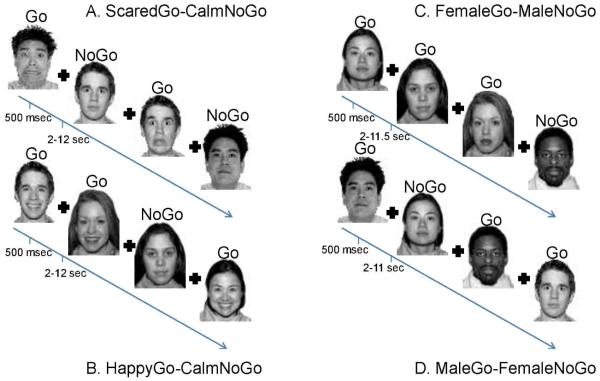
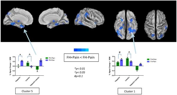
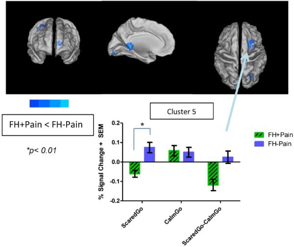
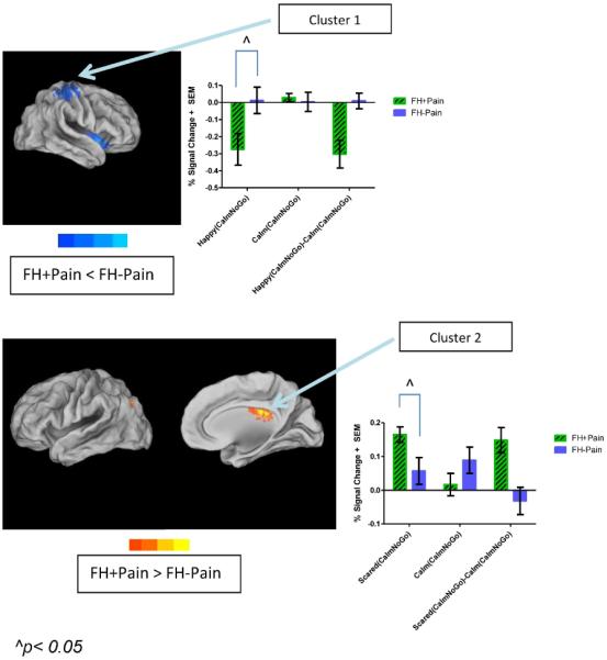
Parental history of chronic pain has been associated with self-reported pain in adolescent offspring. This suggests that there may be neurobiological mechanisms associated with pain heritability. Because emotional circuitry is an important component of pain processing and may also influence cognition, we used functional magnetic resonance imaging to examine affective processing and cognitive control using an Emotional Go/NoGo task in youth with (FH + Pain, n = 8) and without (FH - Pain, n = 8) a parental history of chronic pain (mean age = 14.17 ± .34 years). FH + Pain youth had widespread reductions in brain activity within limbic and visual processing regions during processing of positively valenced emotional stimuli, as well as reduced frontoparietal response while processing negatively valenced emotional stimuli compared with their peers. In addition, during inhibition within a positive emotional context, FH + Pain youth had reduced cognitive control and salience-related brain activity. On the other hand, default mode-related brain response was increased during inhibitory control within a negative emotional context in these adolescents compared with their peers (P/α < .05). The current findings indicate differences in both emotional processing and cognitive control brain response in FH + Pain compared with FH - Pain youth, suggesting that both affective and executive functioning pathways may be important markers related to the intergenerational transmission of pain. Perspective: This is the first study to examine neurobiological markers of pain risk in adolescents with a family history of chronic pain. These findings may aid in the identification of neural phenotypes related to vulnerability for the onset of pain in at-risk youth.

Keywords: Chronic pain; cognitive control; emotion; family history; youth.


Figures

Figure 1
Figure 1. Emotional Go-NoGo Task
Participants completed the Emotional Go-NoGo fMRI task in the scanner. There were four runs of the task: two emotional runs (A and B) and two control runs (C and D). Emotional runs always followed controls runs, but the order in which happy and scared runs appeared was counterbalanced across participants. The task instructions were to respond as quickly and as accurately to the target (go) face that was specified for a particular run and to not respond when a non-target (nogo) face appeared. Each face was presented for 500 milliseconds with a 2-12 second jitter used as the intertrial interval for the emotional runs of the task, and 2-11.5 second jitter used for the control runs of the task. A fixation cross appeared during the jitter period.
Figure 2
Figure 2. Significant differences in brain response to positively valenced faces between FH+Pain and FH−Pain youth
Brain response to Happy vs. Calm Go faces was reduced in FH+Pain youth compared with their FH−Pain peers, as indicated by areas in cool colors surface-mapped on the Population-Average, Landmark- and Surface-based (PALS-B12) template brain, multiple comparison corrected, (p/α<0.05/0.05). These nine clusters are labeled on the maps with names corresponding to regions where the peak coordinate of differences in brain response was located, and include occipital, parietal, frontal, thalamic, and limbic brain regions. Brain activity is bar graphed from two of these clusters (Clusters 1 and 5) as examples of patterns seen in both the contrast of Happy vs. Calm Go faces, and the simple effects of those contrasts. In both regions group differences suggest blunted activity to Happy faces, but increased activity to Calm faces in FH+Pain youth, compared with their FH−Pain peers. L = left, R = right.
Figure 3
Figure 3. Significant differences in brain response to negatively valenced faces between FH+Pain and FH−Pain youth
Brain response to Scared vs. Calm Go faces was reduced in FH+Pain youth compared with their FH−Pain peers, as indicated by areas in cool colors surface-mapped on the Population-Average, Landmark- and Surface-based (PALS-B12) template brain, multiple comparison corrected, (p/α<0.05/0.05). These five clusters are labeled on the maps with names corresponding to regions where the peak coordinate of differences in brain response was located, and include frontal, occipital, parietal, and cingulate cortex. Brain activity is bar graphed from one of these clusters (Cluster 5) as an example of patterns seen in both the contrast of Scared vs. Calm Go faces, and the simple effects of those contrasts. In this region group differences suggest blunted activity to Scared faces, but no significant differences in activity to Calm faces in FH+Pain youth, compared with their FH−Pain peers. L = left, R = right.
Figure 4
Figure 4. Significant differences in brain response during inhibitory control in both positively and negatively valenced contexts between FH+Pain and FH−Pain youth
Brain response to Happy(CalmNoGo) vs. Calm(CalmNoGo) faces was reduced, while brain activity to Scared(CalmNoGo) vs. Calm(CalmNoGo) faces was increased in FH+Pain youth compared with their FH−Pain peers. This is indicated by areas in cool, and warm colors, respectively, surface-mapped on the Population-Average, Landmark- and Surface-based (PALS-B12) template brain, multiple comparison corrected, (p/α<0.05/0.05). All clusters are labeled on the maps with names corresponding to regions where the peak coordinate of differences in brain response was located. In the top panel, this includes the postcentral gyrus and inferior frontal gyrus. Brain activity is bar graphed from two of these clusters (Cluster 1), with simple effects indicating reduced inhibitory control brain response in positively valenced emotional contexts in FH+Pain youth compared with their FH−Pain peers. In the bottom panel, brain activity is increased in middle temporal and cingulate gyri, suggesting reduced suppression of some areas of the default mode network during cognitive control in FH+Pain youth, compared with their FH−Pain peers. L = left, R = right.

References

    1. Anttila P, Metsahonkala L, Helenius H, Sillanpaa M. Predisposing and provoking factors in childhood headache. Headache. 2000;40:351–356. - PubMed
    1. Bair MJ, Poleshuck EL, Wu J, Krebs EK, Damush TM, Tu W, Kroenke K. Anxiety but not social stressors predict 12-month depression and pain severity. Clin J Pain. 2013;29:95–101. - PMC - PubMed
    1. Baliki MN, Chialvo DR, Geha PY, Levy RM, Harden RN, Parrish TB, Apkarian AV. Chronic pain and the emotional brain: specific brain activity associated with spontaneous fluctuations of intensity of chronic back pain. J Neurosci. 2006;26:12165–12173. - PMC - PubMed
    1. Baliki MN, Geha PY, Apkarian AV, Chialvo DR. Beyond feeling: chronic pain hurts the brain, disrupting the default-mode network dynamics. J Neurosci. 2008;28:1398–1403. - PMC - PubMed
    1. Carskadon MA, Acebo C. A self-administered rating scale for pubertal development. J of Adolesc Health. 1993;14:190–195. - PubMed

Publication types

MeSH terms