Review
. 2015 Oct;185(10):2596-606.
doi: 10.1016/j.ajpath.2015.06.001. Epub 2015 Jun 26.

Monocyte and macrophage plasticity in tissue repair and regeneration

Affiliations
Review

Monocyte and macrophage plasticity in tissue repair and regeneration

Amitava Das et al. Am J Pathol. 2015 Oct.

Abstract

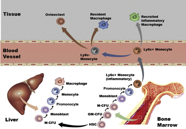
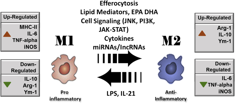
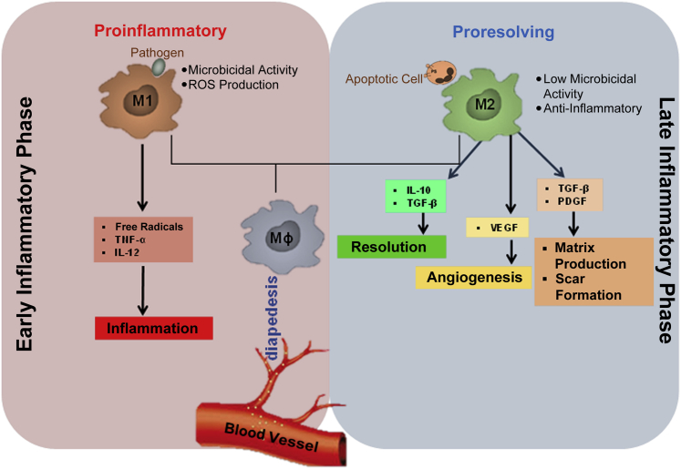
Heterogeneity and high versatility are the characteristic features of the cells of monocyte-macrophage lineage. The mononuclear phagocyte system, derived from the bone marrow progenitor cells, is primarily composed of monocytes, macrophages, and dendritic cells. In regenerative tissues, a central role of monocyte-derived macrophages and paracrine factors secreted by these cells is indisputable. Macrophages are highly plastic cells. On the basis of environmental cues and molecular mediators, these cells differentiate to proinflammatory type I macrophage (M1) or anti-inflammatory or proreparative type II macrophage (M2) phenotypes and transdifferentiate into other cell types. Given a central role in tissue repair and regeneration, the review focuses on the heterogeneity of monocytes and macrophages with current known mechanisms of differentiation and plasticity, including microenvironmental cues and molecular mediators, such as noncoding RNAs.


Figures

Figure 1
Figure 1
Origin and development of the mononuclear phagocyte system. Hematopoietic stem cells (HSCs) in fetal liver or adult bone marrow develop into a progenitor of both macrophages and granulocytes. The granulocyte-macrophage colony-forming unit (GM-CFU) population can commit to the macrophage colony-forming unit (M-CFU) or the granulocyte CFU group of cells. Before becoming macrophages, the M-CFU differentiates into monoblasts, promonocytes, and mature monocytes. This process requires the growth factor colony-stimulating factor-1. In mice, Ly6C is a marker for an inflammatory population of monocytes. In humans, the corresponding marker is CD16.
Figure 2
Figure 2
Macrophage polarization from a proinflammatory M1 to an anti-inflammatory M2 phenotype is mediated by a set of specific factors, including efferocytosis of apoptotic cells present at the site of inflammation, cytokines, lipid mediators, such as eicosapentaenoic acid (EPA) and docosahexaenoic acid (DHA), and cell signaling mediators c-Jun N-terminal kinase (JNK), janus kinase (JAK)–signal transducer and activator of transcription (STAT), and phosphatidylinositol 3-kinase (PI3K). The miRNAs and long noncoding RNAs (lncRNAs) also support macrophage polarization. The expressions of specific genes are altered in transition from M1 to M2. Up-regulated genes are shown with red arrowheads, whereas down-regulated genes are shown with green arrowheads. Arg-1, arginine-1; iNOS, inducible nitric oxide synthase; LPS, lipopolysachharide; MHC-II, major histocompatibility complex class II; TNF-α, tumor necrosis factor α; Ym-1, chitinase 3-like 3.
Figure 3
Figure 3
Macrophage plasticity in tissue repair. After injury, the macrophage (Mϕ) arrives at the site of injury from the systemic circulation via diapedesis. In the early phase, the inflammatory milieu drives macrophage toward M1 polarization. M1 macrophages possess potent microbicidal properties and support IL-12–mediated type 1 helper T-cell responses, which are essential in the early stages of wound healing. In the late inflammatory phase, the change in wound microenvironment and the process of efferocytosis (clearance of apoptotic cells) drive the M1 macrophages toward M2 polarization. M2 supports type 2 helper T-cell–related effector functions and plays a more reparative role in the later stages of wound healing. Macrophages play a major role in the transition of the wounds from the inflammatory to the resolution or proliferative phase, driving angiogenesis and matrix production. PDGF, platelet-derived growth factor; TGF, transforming growth factor; TNF, tumor necrosis factor; VEGF, vascular endothelial growth factor.

References

    1. Sica A., Mantovani A. Macrophage plasticity and polarization: in vivo veritas. J Clin Invest. 2012;122:787–795. - PMC - PubMed
    1. Strauss O., Rod Dunbar P., Bartlett A., Phillips A. The immunophenotype of the antigen presenting cells of the mononuclear phagocyte system in the normal human liver: a systematic review. J Hepatol. 2015;62:458–468. - PubMed
    1. Hume D.A., Ross I.L., Himes S.R., Sasmono R.T., Wells C.A., Ravasi T. The mononuclear phagocyte system revisited. J Leukoc Biol. 2002;72:621–627. - PubMed
    1. Taylor P.R., Gordon S. Monocyte heterogeneity and innate immunity. Immunity. 2003;19:2–4. - PubMed
    1. Hume D.A. Differentiation and heterogeneity in the mononuclear phagocyte system. Mucosal Immunol. 2008;1:432–441. - PubMed

Publication types