Review
. 2015 Jun 29:12:16.
doi: 10.1186/s12987-015-0012-z.

A new glaucoma hypothesis: a role of glymphatic system dysfunction

Affiliations
Review

A new glaucoma hypothesis: a role of glymphatic system dysfunction

Peter Wostyn et al. Fluids Barriers CNS. .

Abstract

In a recent review article titled "A new look at cerebrospinal fluid circulation", Brinker et al. comprehensively described novel insights from molecular and cellular biology as well as neuroimaging research, which indicate that cerebrospinal fluid (CSF) physiology is much more complex than previously believed. The glymphatic system is a recently defined brain-wide paravascular pathway for CSF and interstitial fluid exchange that facilitates efficient clearance of interstitial solutes, including amyloid-β, from the brain. Although further studies are needed to substantiate the functional significance of the glymphatic concept, one implication is that glymphatic pathway dysfunction may contribute to the deficient amyloid-β clearance in Alzheimer's disease. In this paper, we review several lines of evidence suggesting that the glymphatic system may also have potential clinical relevance for the understanding of glaucoma. As a clinically acceptable MRI-based approach to evaluate glymphatic pathway function in humans has recently been developed, a unique opportunity now exists to investigate whether suppression of the glymphatic system contributes to the development of glaucoma. The observation of a dysfunctional glymphatic system in patients with glaucoma would provide support for the hypothesis recently proposed by our group that CSF circulatory dysfunction may play a contributory role in the pathogenesis of glaucomatous damage. This would suggest a new hypothesis for glaucoma, which, just like Alzheimer's disease, might be considered then as an imbalance between production and clearance of neurotoxins, including amyloid-β.


Figures
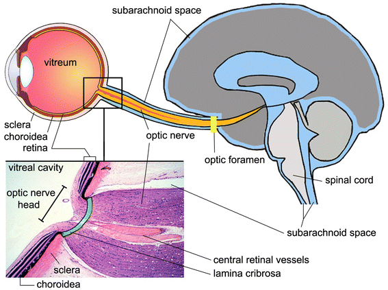
Figure 1
Figure 1
Section of eye, optic nerve head and brain, illustrating the subarachnoid space. The lamina cribrosa (green) is a sieve-like structure in the posterior part of the sclera and allows the passage of the retinal ganglion cell axons and the central retinal vessels. The optic nerve is surrounded by cerebrospinal fluid (blue) in the subarachnoid space. The enlarged histologic view is modified and reproduced with permission, Boston University Histology Learning System (Deborah W. Vaughan, PhD).
Figure 2
Figure 2
Schematic depiction of the glymphatic pathway in the brain. In this brain-wide pathway, cerebrospinal fluid from the subarachnoid space rapidly enters the brain along paravascular channels surrounding penetrating arteries and exchanges with brain interstitial fluid (ISF). ISF and parenchymal solutes (e.g., Aβ) are cleared from the brain along paravenous routes. The green arrow has been added as a reminder that fluid secreted by the blood–brain barrier contributes to the fluid in the parenchyma. Reproduced with permission from Hladky and Barrand [29], their Figure 9a (the glymphatic proposal).

References

    1. Brinker T, Stopa E, Morrison J, Klinge P. A new look at cerebrospinal fluid circulation. Fluids Barriers CNS. 2014;11:10. doi: 10.1186/2045-8118-11-10. - DOI - PMC - PubMed
    1. Oresković D, Klarica M. The formation of cerebrospinal fluid: nearly a hundred years of interpretations and misinterpretations. Brain Res Rev. 2010;64:241–262. doi: 10.1016/j.brainresrev.2010.04.006. - DOI - PubMed
    1. Igarashi H, Tsujita M, Kwee IL, Nakada T. Water influx into cerebrospinal fluid is primarily controlled by aquaporin-4, not by aquaporin-1: 17O JJVCPE MRI study in knockout mice. NeuroReport. 2014;25:39–43. - PMC - PubMed
    1. Iliff JJ, Wang M, Liao Y, Plogg BA, Peng W, Gundersen GA, et al. A paravascular pathway facilitates CSF flow through the brain parenchyma and the clearance of interstitial solutes, including amyloid β. Sci Transl Med. 2012;4:147ra111. doi: 10.1126/scitranslmed.3003748. - DOI - PMC - PubMed
    1. Yang L, Kress BT, Weber HJ, Thiyagarajan M, Wang B, Deane R, et al. Evaluating glymphatic pathway function utilizing clinically relevant intrathecal infusion of CSF tracer. J Transl Med. 2013;11:107. doi: 10.1186/1479-5876-11-107. - DOI - PMC - PubMed

Publication types