Case Reports
. 2015 Jul;103(7):630-40.
doi: 10.1002/bdra.23397. Epub 2015 Jun 27.

EFTUD2 deficiency in vertebrates: Identification of a novel human mutation and generation of a zebrafish model

Affiliations
Case Reports

EFTUD2 deficiency in vertebrates: Identification of a novel human mutation and generation of a zebrafish model

Brett Deml et al. Birth Defects Res A Clin Mol Teratol. 2015 Jul.

Abstract

Background: Congenital microphthalmia and coloboma are severe developmental defects that are frequently associated with additional systemic anomalies and display a high level of genetic heterogeneity.

Methods: To identify the pathogenic variant in a patient with microphthalmia, coloboma, retinal dystrophy, microcephaly, and other features, whole exome sequencing analysis of the patient and parental samples was undertaken. To further explore the identified variant/gene, expression and functional studies in zebrafish were performed.

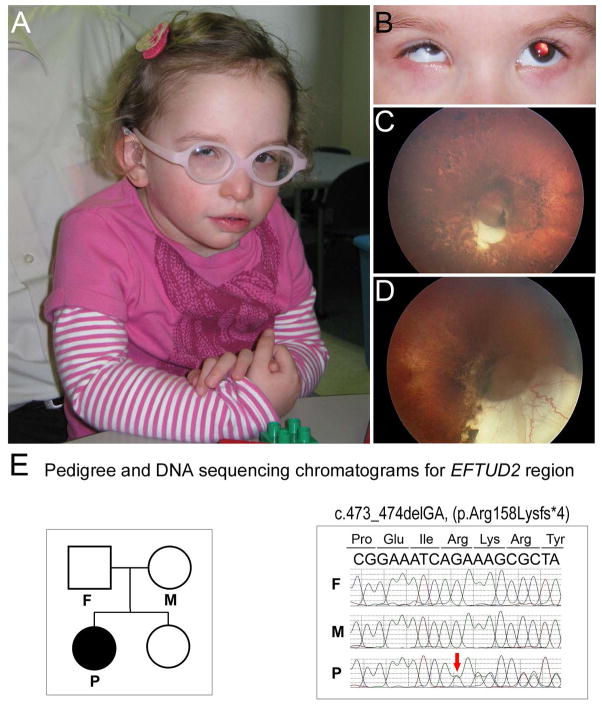
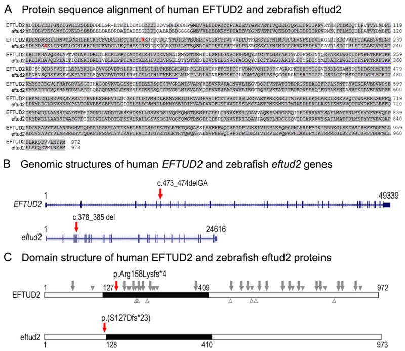
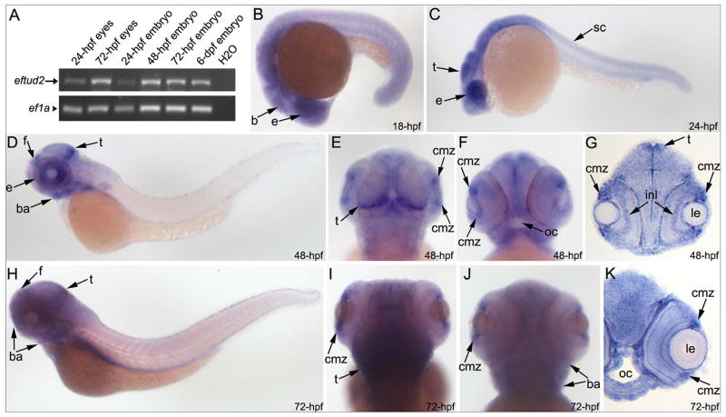
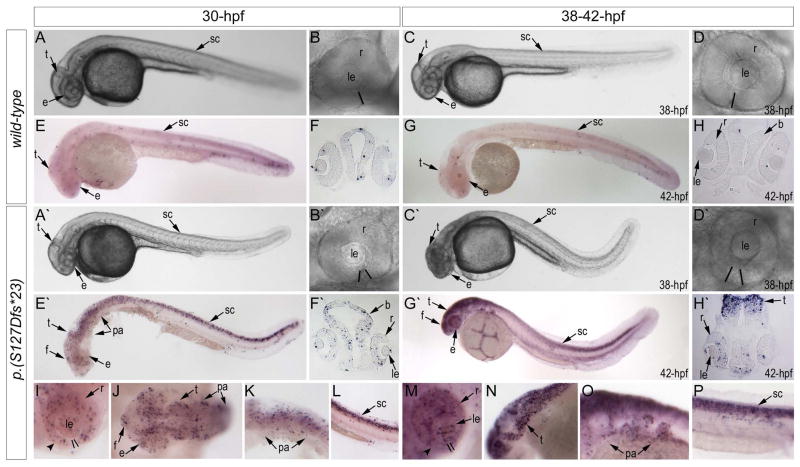
Results: Whole exome sequencing revealed a de novo variant, c.473_474delGA, p.(Arg158Lysfs*4), in EFTUD2 which encodes a component of the spliceosome complex. Dominant mutations in EFTUD2 cause Mandibulofacial Dysostosis, Guion-Almeida type, which does not involve microphthalmia, coloboma, or retinal dystrophy; analysis of genes known to cause these ocular phenotypes identified several variants of unknown significance but no causal alleles in the affected patient. Zebrafish eftud2 demonstrated high sequence conservation with the human gene and broad embryonic expression. TALEN-mediated disruption was employed to generate a c.378_385 del, p.(Ser127Aspfs*23) truncation mutation in eftud2. Homozygous mutants displayed a reduced head size, small eye, curved body, and early embryonic lethality. Apoptosis assays demonstrated a striking increase in terminal deoxynucleotidyl transferase-mediated deoxyuridine triphosphate nick end-labeling (TUNEL)-positive cells in the developing brain, eye, spinal cord, and other tissues starting at 30 hours postfertilization.

Conclusion: This study reports a novel mutation in EFTUD2 in a Mandibulofacial Dysostosis, Guion-Almeida type patient with unusual ocular features and the generation of a first animal model of eftud2 deficiency. The severe embryonic phenotype observed in eftud2 mutants indicates an important conserved role during development of diverse tissues in vertebrates.

Keywords: EFTUD2; coloboma; microphthalmia; retinal dystrophy; zebrafish.


Conflict of interest statement

The authors declare no competing financial interests.

Figures

Figure 1
Figure 1. Clinical images and EFTUD2 sequence of Patient 1
A–D: Patient images. Please note craniofacial features of microcephaly, low-set crumpled ears, broad nasal bridge, upsplanting palpebral fissures, left epicanthic fold, mandibular and malar hypoplasia, triangular facies, and mild facial asymmetry as well as microphthalmia/microcornea (A–B). Retinal images demonstrate moderately sized optic pit with inferior cresent, lacunar ‘punched out’ defects with hyper- and hypo- pigmentation scattered in a ‘bird-shot’ fashion throughout the entire retina, attenuated vessels, and hypoplastic macula in the left eye (C) and large optic pit and inferior coloboma, severely attenuated/absent retinal vasculature, and a mild peripheral pigmentary dystrophy in the right eye (D). E. Pedigree (on the left) and DNA sequencing chromatograms (on the right). Individuals included in trio analysis are indicated on the pedigree; affected proband indicated with black symbol, P, proband, M, mother, F, father. Chromatogram shows the presence of the frameshift variant (red arrow) in the proband and its absence in both parents.
Figure 2
Figure 2. Comparison of human and zebrafish EFTUD2/eftud2 genomic and protein structure
A. Protein sequence alignment of human EFTUD2 and zebrafish eftud2; amino acids identical between the homologs are highlighted with a light grey color, the amino acids representing the beginning of the frameshift in the human patient and zebrafish line are indicated in red font in the respective protein. The blue underline indicates the amino acids involved in the translational (tr)-type guanine nucleotide-binding (G) domain. B. Genomic structures of the human EFTUD2 and zebrafish eftud2 genes as indicated in the Genome Browser for the respective genes. The exons are shown as blue boxes with 5′ and 3′UTR shown as short and coding regions shown as tall blue boxes. The positions of the human and zebrafish variants are indicated with red arrows on the respective genes. C. Domain structure of human EFTUD2 and zebrafish eftud2 proteins. The black box shows the translational (tr)-type guanine nucleotide-binding (G) domain; the positions of the human and zebrafish variants that were identified/generated in this study are indicated with red arrows on the respective proteins; the approximate positions of the unique previously reported pathogenic variants are shown on the human protein with truncating variants indicated above as grey arrows (nonsense and frameshift) or solid grey arrowheads (splicing variants, indicated based on affected exon) above and missense variants indicated as empty grey arrowheads below.
Figure 3
Figure 3. Embryonic expression of zebrafish eftud2
RT-PCR and in situ hybridization expression data for eftud2 in zebrafish embryos. Please note the presence of eftud2 transcript in 24- and 72-hpf embryonic eyes as well as whole embryos as detected by RT-PCR (A). In situ hybridization revealed a broad expression throughout the embryo with a possible enrichment in the head and eye region. Whole mount images (B–F, H–J) and sections (G, K) are shown; developmental stage is indicated in the lower-right corner; b- brain, ba- branchial arches, cmz-ciliary marginal zone of the retina, e-eye, f- forebrain, inl- inner nuclear layer of the retina; le- lens, oc-oral cavity, sc- spinal cord, t-tectum.
Figure 4
Figure 4. Phenotypic analysis of eftud2 mutants
A–H. Morphology (AD) and TUNEL staining (E–H) of wild-type embryos at 30 and 38–42-hpf (as indicated). A–H′. Morphology (A′–D′) and TUNEL staining (E′–H′) of eftud2 mutant embryos at 30 and 38–42-hpf. Please note microcephaly and coloboma in mutant embryos. I–P. Enlarged whole mount images of TUNEL staining in eftud2 mutant embryos in representative embryonic regions. Please note increase in TUNEL positive cells throughout the embryo, particularly in the eye, branchial arches, tectum, and spinal cord. The black lines in B, B′, D, D′, I and M indicate the retinal edges and highlight the presence of coloboma in mutant embryos; arrowheads in I and M indicate thinning of the ventral portion of the retina in mutants; b- brain, e-eye, f- forebrain, le- lens, pa- pharyngeal arches, r-retina, sc- spinal cord, t-tectum.

References

    1. Aanes H, Ostrup O, Andersen IS, Moen LF, Mathavan S, Collas P, Alestrom P. Differential transcript isoform usage pre- and post-zygotic genome activation in zebrafish. BMC Genomics. 2013;14 331-2164-14-331. - PMC - PubMed
    1. Bardakjian TM, Schneider A. The genetics of anophthalmia and microphthalmia. Curr Opin Ophthalmol. 2011;22:309–313. - PubMed
    1. Beleggia F, Li Y, Fan J, Elcioglu NH, Toker E, Wieland T, Maumenee IH, Akarsu NA, Meitinger T, Strom T, Lang R, Wollnik B. CRIM1 haploinsufficiency causes defects in eye development in human and mouse. Hum Mol Genet 2015 - PMC - PubMed
    1. Benaglio P, San Jose PF, Avila-Fernandez A, Ascari G, Harper S, Manes G, Ayuso C, Hamel C, Berson EL, Rivolta C. Mutational screening of splicing factor genes in cases with autosomal dominant retinitis pigmentosa. Mol Vis. 2014;20:843–851. - PMC - PubMed
    1. Bernier FP, Caluseriu O, Ng S, Schwartzentruber J, Buckingham KJ, Innes AM, Jabs EW, Innis JW, Schuette JL, Gorski JL, Byers PH, Andelfinger G, Siu V, Lauzon J, Fernandez BA, McMillin M, Scott RH, Racher H, Majewski J, Nickerson DA, Shendure J, Bamshad MJ, Parboosingh JS FORGE Canada Consortium. Haploinsufficiency of SF3B4, a component of the pre-mRNA spliceosomal complex, causes Nager syndrome. Am J Hum Genet. 2012;90:925–933. - PMC - PubMed

Publication types

Substances

LinkOut - more resources