. 2015 Jul 15;87(2):341-54.
doi: 10.1016/j.neuron.2015.06.007. Epub 2015 Jun 25.

Silencing Nociceptor Neurons Reduces Allergic Airway Inflammation

Affiliations

Silencing Nociceptor Neurons Reduces Allergic Airway Inflammation

Sébastien Talbot et al. Neuron. .

Abstract

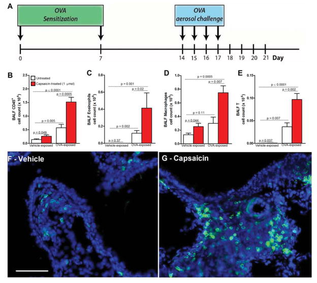
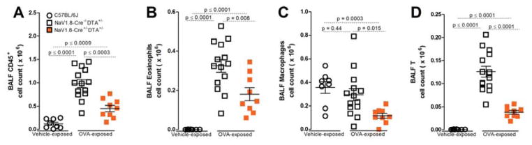
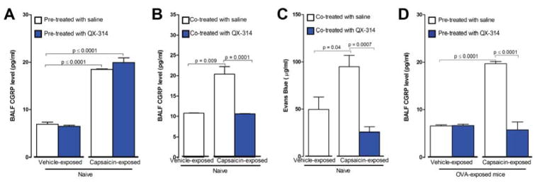
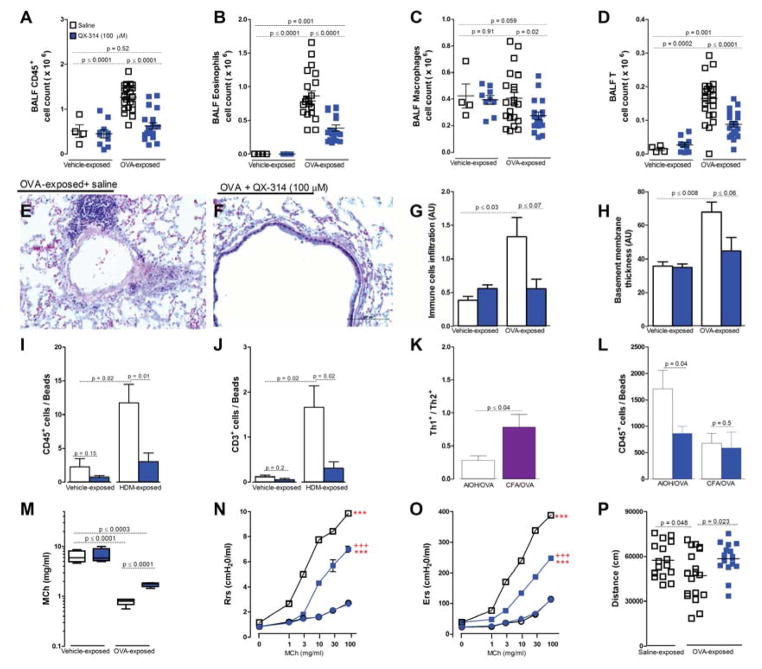
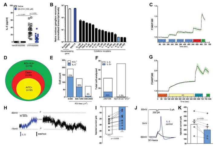
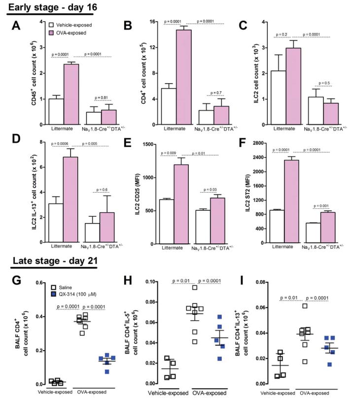
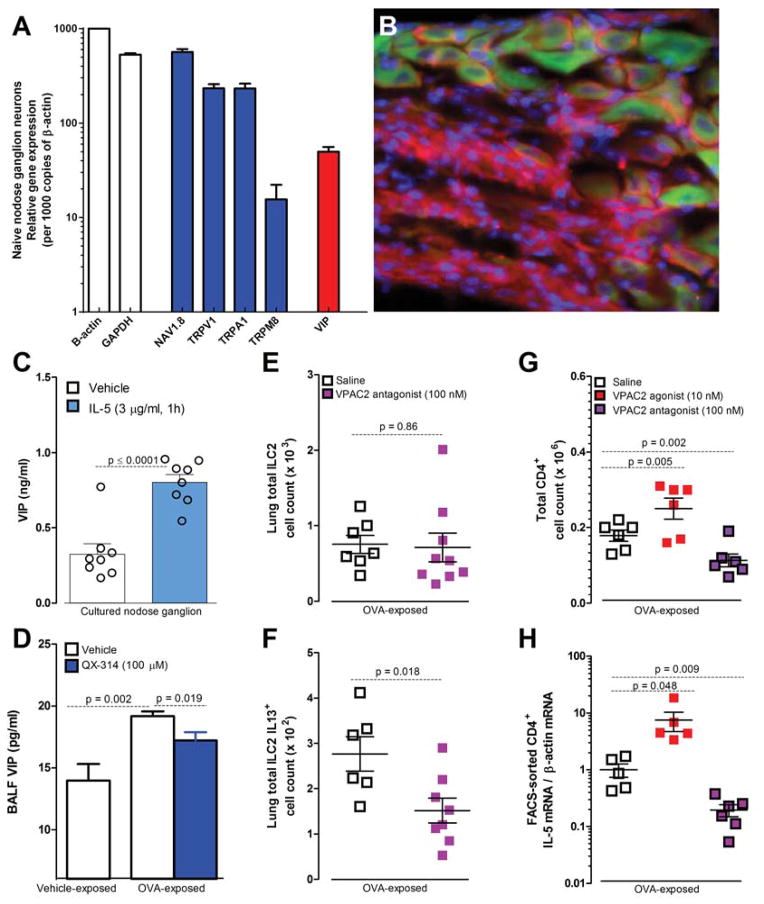
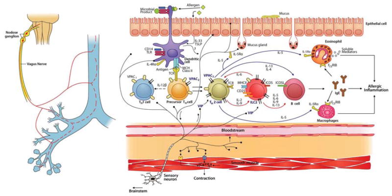
Lung nociceptors initiate cough and bronchoconstriction. To elucidate if these fibers also contribute to allergic airway inflammation, we stimulated lung nociceptors with capsaicin and observed increased neuropeptide release and immune cell infiltration. In contrast, ablating Nav1.8(+) sensory neurons or silencing them with QX-314, a charged sodium channel inhibitor that enters via large-pore ion channels to specifically block nociceptors, substantially reduced ovalbumin- or house-dust-mite-induced airway inflammation and bronchial hyperresponsiveness. We also discovered that IL-5, a cytokine produced by activated immune cells, acts directly on nociceptors to induce the release of vasoactive intestinal peptide (VIP). VIP then stimulates CD4(+) and resident innate lymphoid type 2 cells, creating an inflammatory signaling loop that promotes allergic inflammation. Our results indicate that nociceptors amplify pathological adaptive immune responses and that silencing these neurons with QX-314 interrupts this neuro-immune interplay, revealing a potential new therapeutic strategy for asthma.


Conflict of interest statement

The authors have declared that no conflicts of interest exist.

Figures

Figure 1
Figure 1. Nociceptor activation promotes airway inflammation
(A) Inflammation induced in mice following i.p. allergen (ovalbumin (OVA)) sensitization (day 0 and 7, with AlOH) and inhaled challenge (days 14–17). Drugs/vehicle administered at peak of OVA-induced inflammation (day 18) and impact assessed on day 21. Capsaicin (B–E; 1 μmol, red bar) or vehicle (white bar) instilled (intranasally) on Day 17 after vehicle or OVA challenge (24h prior to BALF measurement). Capsaicin increased airway CD45+ (total immune) (B), eosinophil (C), macrophage (D) and lymphocyte (E) cell counts. T-cell influx (green, LCK-Cre+/−eGFP+/−) in control lungs following vehicle (F) or capsaicin (G; 1 μmol) instillation. Scale bar 10 μm. Mean ± S.E.M; Two-tailed unpaired Student’s t-test (n = 6–10 animals/group; 2 cohorts).
Figure 2
Figure 2. Lung sensory neuron ablation reduces airway allergic inflammation
Four days after last aerosol challenge (day 21), OVA-exposed mice developed CD45+ cell count increases in BALF (A), including eosinophils (B) and lymphocytes (D), but not macrophages (C). Ablation of NaV1.8 (NaV1.8+/− DTA+/−, orange squares; A–D) expressing sensory neurons significantly decreased these levels. Mean ± S.E.M; Two-tailed unpaired Student’s t-test (n = 4–12 animals/group; 1–2 cohorts).
Figure 3
Figure 3. Airway sensory neuron silencing with QX-314 abolishes capsaicin-induced peptide release and vascular leak
To assess nociceptor-mediated peptide release capsaicin (1 μmol, intranasal) was administered to naïve mice. BALF CGRP levels were increased one hour following the capsaicin-challenge (A–B). QX-314 (100 μM) treatment 1 hr prior to the capsaicin did not alter capsaicin-induced iCGRP levels (A) but QX-314 (100 μM) when administered immediately after the capsaicin, blocked the CGRP rise (B) (n = 4–15 animals/group; 2–3 cohorts). Reduction in vascular leak (C) following co-administration of capsaicin (1 μmol) with QX-314 (100 μM) (n = 4–5 animals/group; 1 cohort). BALF CGRP levels increased following capsaicin (1 μmol) instillation in OVA-challenged mice (D) and this was blocked by QX-314 pre-treatment one hour before (100 μM). Mean ± S.E.M; Two-tailed unpaired Student’s t-test (n = 5–12 animals/group; 2 cohorts).
Figure 4
Figure 4. Airway sensory neuron silencing reduces lung inflammation and hyperresponsiveness
OVA-exposed mice develop increased CD45+ immune cell counts in BALF (A), that include eosinophils (B) and lymphocytes (D), but not macrophages (C). Silencing lung sensory neurons with QX-314 (100 μM, 72h prior to measurement, blue squares) decreased these immune cell responses (A–H). Representative Hematoxylin and Eosin (H&E)-stained sections of OVA exposed lungs treated with saline (E) or QX-314 (100 μM; F). Scale 100 μm. Immune cell infiltration severity (G) and basement membrane thickening (H) measured by tissue morphometry in HE- sections. House dust mite (HDM) challenge increased BALF CD45+ (I) and lymphocyte counts (J), an effect reversed by QX-314 (100 μM). CFA/OVA sensitization produced a greater increase in Th1/Th2 cell ratio than AlOH/OVA sensitization (K). QX-314 (100 μM) reduced BALF CD45+ cell counts in the AlOH/OVA but not CFA/OVA sensitized model (L). Mean ± S.E.M; Two-tailed unpaired Student’s t-test (n = 4–25 animals/group; 4 cohorts). Change in airway reactivity measured as ED200RL (M), Resistance (N) Elastance (O) in OVA-exposed mice treated with QX-314 (D–F; 100 μM, day 18) or saline. Difference vehicle- (***) and OVA-exposed (+++) groups P <0.001. OVA-exposed mice performed less voluntary wheel running (P) (1h assessment) than control mice, an effect reversed by QX-314 (100 μM, day 18) assessed 24h after silencing. Two-tailed unpaired Student’s t-test (n = 4–40 animals/group; 2–3 cohorts).
Figure 5
Figure 5. IL-5 activates NaV1.8+ nodose ganglion neurons
IL-5 levels in BALFs increased in OVA challenged mice, an effect reversed by QX-314 (100 μM) (A). Transcript profiling of naïve nodose ganglia reveals basal expression of cytokine receptors including IL-5R (B). Cultured nodose ganglion neurons from OVA-exposed mice show a dose-dependent calcium increase (C) in response to IL-5 (0.3–10 μg/ml). Venn diagram shows overlapping populations of KCL+IL-5+ and AITC or capsaicin responsive neurons (D). IL-5 responsive cells are mainly small and medium size (E) and nearly null in OVA-exposed Nav1.8-Cre +/− DTA+/− mice (F). IL5 mediated calcium increase absent in 0 external Ca2+ (G). Current-clamp recordings of small cells (capacitance <45 pF) showing that IL-5 (1 μg/ml) depolarizing resting membrane potential (RMP) by ~6 mV (H, I; n = 12 cells). The current needed to trigger an action potential (rheobase) is smaller when IL-5 is present (J, K; n = 7 cells). Mean ± S.E.M; Two-tailed unpaired (A–G) and paired (H–K) Student’s t-test (n = 3 biological replicate per group).
Figure 6
Figure 6. Sensory neurons control ILC2 and CD4+ cell recruitment and activation in the lung
On day 16 (A–F), OVA-exposed mice (pink bars) with intact nociceptors (littermate; NaV1.8−/−DTA+/−) had higher whole lung CD45+ (A), CD4+ (B), ILC2 (C), ILC2 IL-13+ cell counts (D), as well as ILC2 CD25 (E) and ST2 mean fluorescence intensity MFI) (F) than mice with ablated nociceptors (NaV1.8+/−DTA+/−). QX-314 treatment (100 μM, day 18; blue squares) decreased OVA-induced CD4+ cells on day 21 (G), including CD4+IL5+ (H) and CD4+IL13+ (I) cells, relative to control mice (white squares). Mean ± S.E.M; Two-tailed unpaired student’s t-test (n = 4–9 animals/group; 1–2 cohorts).
Figure 7
Figure 7. IL-5 provokes sensory neuron release of VIP to activate ILC2 and recruit CD4+ cells
Gene profiling of naïve NaV1.8+ nodose ganglion neurons reveals expression of nociceptor markers and VIP (A). A VIP reporter mouse (B, VIP-Cre+/−EGFP/td-tomato+/− green) reveals that ~80% of NaV1.8+ (magenta) nodose ganglion neurons express VIP (green) (scale bar = 50 μm) (B). IL-5 (1h, 3 ug/ml) induced VIP release from nodose ganglion neurons from OVA-exposed mice (C). These mice have elevated BALF VIP levels on day 21 compared to vehicle-exposed mice (white bar), which is decreased by QX-314 treatment (100 μM, day 18; blue bar) (D). In OVA-exposed lungs, the VPAC2 antagonist PG 99465 (100 nM, every 12h for 48h starting on day 16; pink squares) did not impact resident ILC2 numbers (E) but reduced ILC2activation; IL-13+ cells (F). The VPAC2 agonist BAY 55-9837 (10nM, every 12h for 96h; red squares) increased, and the VPAC2 antagonist PG 99465 (100 nM, every 12h for 96h; pink squares) decreased CD4+ cell numbers(G) and IL-5 mRNA expression in FACS-sorted CD4+ cells (H). Mean ± S.E.M; Two-tailed unpaired Student’s t-test (n = 3–14 animals/group; 1–2 cohorts).
Figure 8
Figure 8. Model of nociceptor involvement in type-2 inflammation
During allergen exposure, lung afferents are activated by dendritic and epithelial cells. Dendritic cells also polarize precursor TH cells into TH2 cells. Activated nociceptors release VIP, which stimulates lung resident ILC2 and newly differentiated TH2 cells via the VPAC2 receptor. Type 2 cytokines, including IL-5 and IL-13, are released by ILC2 and TH2 cells and initiate the chemotaxis and activation of eosinophils and macrophages, IgE secretion by B cells, mucus production by goblet cells, and smooth muscle contraction, culminating in allergic inflammation and bronchial hyperresponsiveness. IL-5 activates nociceptors to trigger VIP and other neuropeptide release leading to additional IL-5 production. Scheme inspired by Licona-Limon et al., 2013; Vercelli, 2008.

Comment in

References

    1. Akinbami L. The state of childhood asthma, United States, 1980–2005. Adv Data. 2006:1–24. - PubMed
    1. Ansel JC, Brown JR, Payan DG, Brown MA. Substance P selectively activates TNF-alpha gene expression in murine mast cells. J Immunol. 1993;150:4478–4485. - PubMed
    1. Barnes PJ. Neuroeffector mechanisms: the interface between inflammation and neuronal responses. J Allergy Clin Immunol. 1996;98:S73–81. discussion S81–73. - PubMed
    1. Barnes PJ. New drugs for asthma. Nat Rev Drug Discov. 2004;3:831–844. - PubMed
    1. Barnig C, Cernadas M, Dutile S, Liu X, Perrella MA, Kazani S, Wechsler ME, Israel E, Levy BD. Lipoxin A4 regulates natural killer cell and type 2 innate lymphoid cell activation in asthma. Sci Transl Med. 2013;5:174ra126. - PMC - PubMed

Publication types

MeSH terms