. 2015 Jun 29:5:11765.
doi: 10.1038/srep11765.

Stimulation of EphB2 attenuates tau phosphorylation through PI3K/Akt-mediated inactivation of glycogen synthase kinase-3β

Affiliations

Stimulation of EphB2 attenuates tau phosphorylation through PI3K/Akt-mediated inactivation of glycogen synthase kinase-3β

Jun Jiang et al. Sci Rep. .

Abstract

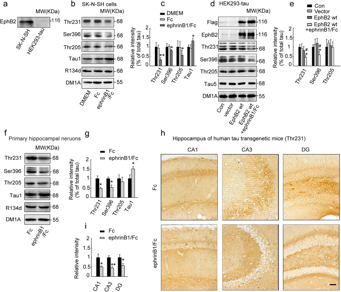
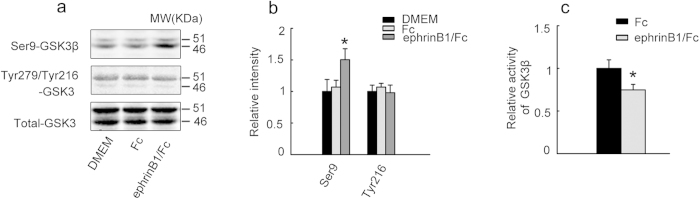
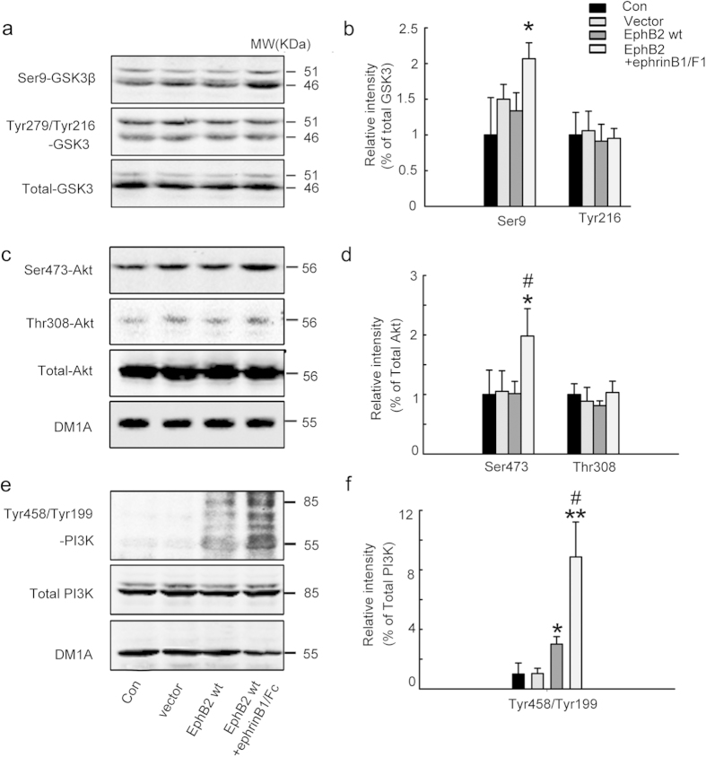
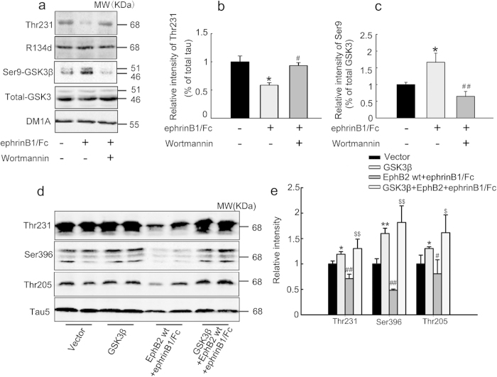
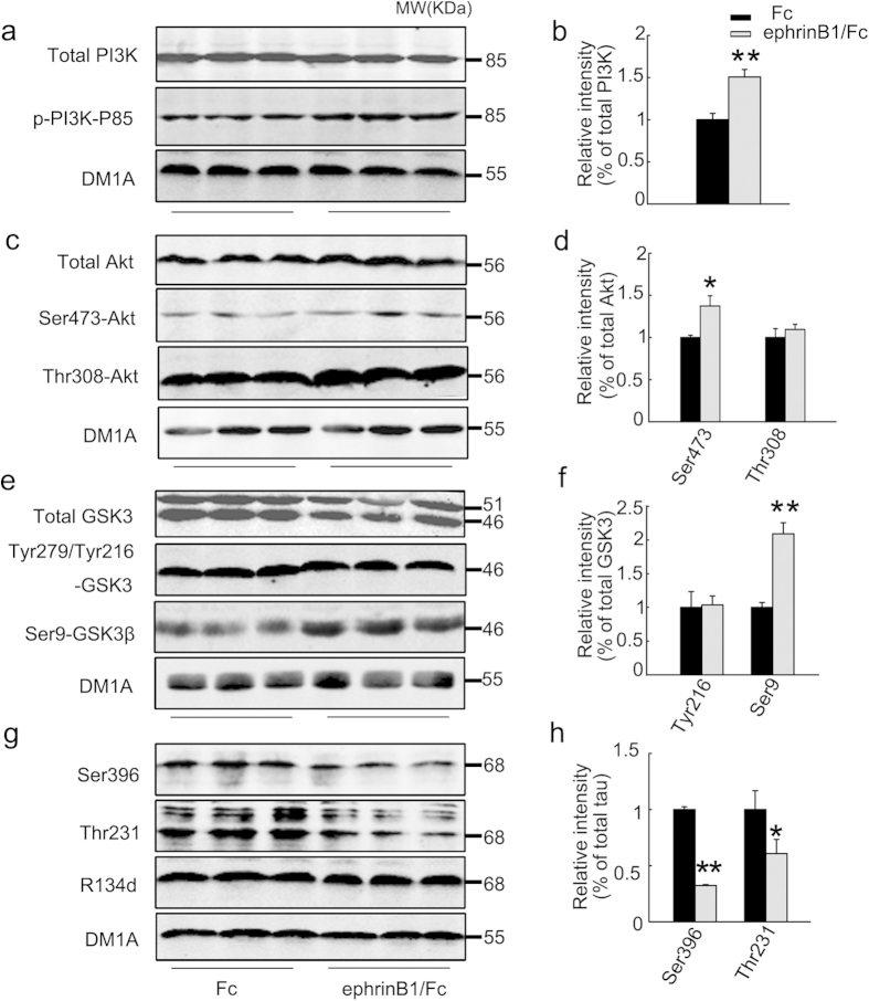
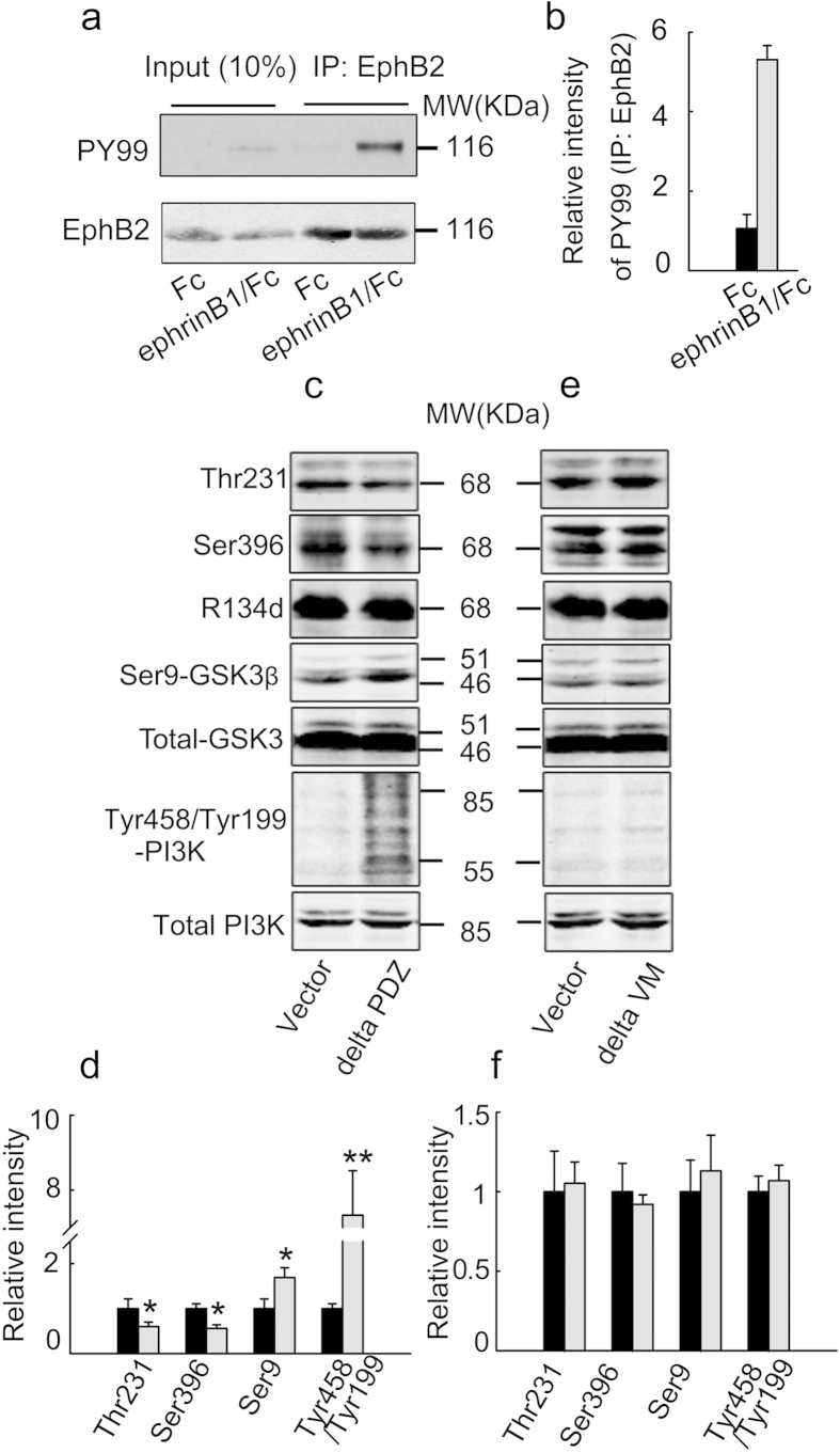
Abnormal tau hyperphosphorylation is an early pathological marker of Alzheimer's disease (AD), however, the upstream factors that regulate tau phosphorylation are not illustrated and there is no efficient strategy to arrest tau hyperphosphorylation. Here, we find that activation of endogenous EphB2 receptor by ligand stimulation (ephrinB1/Fc) or by ectopic expression of EphB2 plus the ligand stimulation induces a remarkable tau dephosphorylation at multiple AD-associated sites in SK-N-SH cells and human embryonic kidney cells that stably express human tau (HEK293-tau). In cultured hippocampal neurons and the hippocampus of human tau transgenic mice, dephosphorylation of tau proteins was also detected by stimulation of EphB2 receptor. EphB2 activation inhibits glycogen synthase kinase-3β (GSK-3β), a crucial tau kinase, and activates phosphatidylinositol-3-kinase (PI3K)/Akt both in vitro and in vivo, whereas simultaneous inhibition of PI3K or upregulation of GSK-3β abolishes the EphB2 stimulation-induced tau dephosphorylation. Finally, we confirm that ephrinB1/Fc treatment induces tyrosine phosphorylation (activation) of EphB2, while deletion of the tyrosine kinase domain (VM) of EphB2 eliminates the receptor stimulation-induced GSK-3β inhibition and tau dephosphorylation. We conclude that activation of EphB2 receptor kinase arrests tau hyperphosphorylation through PI3K-/Akt-mediated GSK-3β inhibition. Our data provide a novel membranous target to antagonize AD-like tau pathology.


Conflict of interest statement

The authors declare no competing financial interests.

Figures

Figure 1
Figure 1. Stimulation of EphB2 induces tau dephosphorylation in cells and hippocampus of human tau transgenic mice.
(a) SK-N-SH but not HEK293-tau cells express endogenous EphB2 receptor measured by Western blotting. (b, c) EphB2 was stimulated by treatment of the SK-N-SH cells with ephrinB1/Fc dissolved in DMEM for 45 min, and then tau phosphorylation at different sites as labeled was measured by Western blotting. (d, e) The flag-labeled wild type EphB2 (EphB2wt) plasmid was transfected into HEK293-tau cells for 24 h and then the cells were treated with ephrinB1/Fc for 45 min before detection of tau phosphorylation. (f, g) The primary hippocampal neurons (cultured for 7 days in vitro) were treated with ephrinB1/Fc for 45 min, and then tau phosphorylation was measured by Western blotting. (h, i) The ephrinB1/Fc was infused into hippocampal CA3 region of the human tau transgenic mice (10 m old) for 45 min, and then tau phosphorylation in hippocampus was measured by immunohistochemistry staining (h) and quantitative analysis (i). Scale bar = 20 μm. Dephosphorylation of tau was detected by stimulation of EphB2 receptor. The total tau was probed by R134d or Tau5 that was normalized against tubulin (DM1A), and the phosphorylation level of tau at Thr205, Thr231, Ser396 and tau-1 sites was normalized against total tau. [Note that tau-1 reacts with the unphosphorylated tau at Ser198/199/202, therefore an increased immunoreaction to tau-1 suggests an increased tau dephosphorylation]. The blots were representative of at least three independent experiments with cells from 9 ~ 12 culture wells for each group. The images were representative of at least 3 mice. *P < 0.05, **P < 0.01 versus Fc or DMEM or vector; #P < 0.05 versus EphB2 wt alone (mean±SD).
Figure 2
Figure 2. Stimulation of EphB2 inhibits GSK-3β.
(a, b) SK-N-SH cells were treated with ephrinB1/Fc or Fc or DMEM (vehicle control) for 45 min to activate EphB2 and then the activity-dependent phosphorylation of GSK-3β at Ser9 (inactivation) and Tyr216 (activation) was measured by Western blotting (a) and quantitative analysis (b). (c) The inactivation of GSK-3β was also observed by biochemical assay of the kinase activity. Data were representative of at least three independent experiments with cells from 9~12 culture wells for each group and presented as the mean±SD. *P < 0.05 versus Fc or DMEM.
Figure 3
Figure 3. Stimulation of EphB2 up-regulates PI3K and Akt with GSK-3β inhibition.
HEK293-tau cells were untreated (Con) or transfected with pcDNA3.0 (Vector) or transfected with wild type EphB2 alone (EphB2wt, inactive) for 24 h, or transfected with EphB2wt for 24 h and then stimulated with ephrinB1/Fc for 45 min. The activity-dependent phosphorylation of GSK-3β at Ser9 (inactive form) and Tyr216 (active form) (a, b), Akt at Ser473 and Thr308 (active form) (c, d), and PI3K at Tyr458/199 (active form) (e, f) was measured respectively by Western blotting and quantitative analysis. The phosphorylation level of the kinases was normalized against the total level. Data were representative of at least three independent experiments with cells from 9~12 culture wells for each group and presented as the mean ± SD. *P < 0.05, **P < 0.01 versus Con and Vector; #P <0.05 versus EphB2 wt.
Figure 4
Figure 4. Simultaneous inhibition of PI3K or overexpression of GSK-3β abolishes the EphB2 activation-induced tau dephosphorylation.
(a-c) SK-N-SH cells were treated with ephrinB1/Fc alone or ephrinB1/Fc plus wortmannin (an inhibitor of PI3K) for 2 h, and then phosphorylation level of GSK-3β at Ser9 and tau at Thr231 was measured by Western blotting. *P < 0.05 versus untreated control; #P < 0.05, ##P < 0.01 versus ephrinB1/Fc. (d, e) HEK293 cells were transfected with pcDNA3.0 (Vector), or with GSK-3β, or with EphB2wt for 24 h and ephrinB1/Fc stimulation for 45 min, or with co-transfection of EphB2wt and GSK-3β□plus ephrinB1/Fc stimulation. The phosphorylation levels of GSK-3β at Ser9 and tau at Thr205, Thr231 and Ser396 were measured by Western blotting. The phosphorylation level of GSK-3β at Ser9 was normalized against total GSK-3β, while the phosphorylation of tau was normalized against total tau probed by R134d or Tau5. Data were representative of at least three independent experiments with cells from 9 ~ 12 culture wells for each group and presented as the mean ± SD. *P < 0.05, **P < 0.01 GSK-3β versus Vector; #P < 0.05, ##P < 0.01 EphB2/ephrinB1/Fc versus GSK-3β; $ <0.05, $$ P < 0.01 GSK-3β/EphB2/ephrinB1/Fc versus EphB2/ephrinB1/Fc.
Figure 5
Figure 5. Stimulation of EphB2 induces tau dephosphorylation with activation of PI3K/Akt and inhibition of GSK-3 in hippocampus of human tau transgenic mice.
EphrinB1/Fc or Fc was infused stereotactically into the hippocampal CA3 region of the human tau transgenic mice for 45 min and then the total and the activity-dependent phosphorylation levels of PI3K (a, b), Akt (c, d), GSK-3 (e, f), and the phosphorylation level of tau (g, h) were measured by Western blotting. The phosphorylation level of the kinases was normalized against total kinase level, while the phosphorylation level of tau was normalized against total tau probed by R134d. The total levels of the kinases and tau were normalized against tubulin probed by DM1A. The blots were representative of at least three independent experiments from at least 6 mice for each group and presented as the mean ± SD. *P < 0.05, **P < 0.01 versus Fc.
Figure 6
Figure 6. EphrinB1/Fc stimulates tyrosine phosphorylation of EphB2 and deletion of the kinase domain (VM) but not the PDZ binding domain eliminates EphB2-induced tau dephosphorylation.
HEK293-tau cells were treated with ephrinB1/Fc for 45 min, and then the phosphorylation level of EphB2 was measured by immunoprecipitation with anti-EphB2 antibody and Western blotting probed by PY99 and anti-EphB2 antibody (a, b). HEK293-tau cells were transfected with deletion of PDZ (c, d) or VM (e, f) domain of EphB2 or the control Vector for 24 h and then treated with ephrinB1/Fc for 45 min. The phosphorylation levels of tau at Thr231 and Ser396, or GSK-3β at Ser9, or PI3K at Tyr458/Tyr199 were measured by Western blotting. The phosphorylation levels of tau and the kinases were normalized against the total levels. Data were representative of at least three independent experiments from 9 ~ 12 culture wells for each group and presented as the mean ± SD. *P < 0.05, **P <0.01 versus Vector.

References

    1. Braak H. & Del Tredici K. The pathological process underlying Alzheimer’s disease in individuals under thirty. Acta neuropathologica 121, 171–181 (2011). - PubMed
    1. Lee V. M., Balin B. J., Otvos L. Jr. & Trojanowski J. Q. A68: a major subunit of paired helical filaments and derivatized forms of normal Tau. Science 251, 675–678 (1991). - PubMed
    1. Grundke-Iqbal I. et al. Abnormal phosphorylation of the microtubule-associated protein tau (tau) in Alzheimer cytoskeletal pathology. Proc Natl Acad Sci USA. 83, 4913–4917 (1986). - PMC - PubMed
    1. Alonso A. C., Zaidi T., Grundke-Iqbal I. & Iqbal K. Role of abnormally phosphorylated tau in the breakdown of microtubules in Alzheimer disease. Proc Natl Acad Sci USA. 91, 5562–5566 (1994). - PMC - PubMed
    1. Billingsley M. L. & Kincaid R. L. Regulated phosphorylation and dephosphorylation of tau protein: effects on microtubule interaction, intracellular trafficking and neurodegeneration. Biochem J. 323 (Pt 3), 577–591 (1997). - PMC - PubMed

Publication types

MeSH terms