. 2015 Jul 7;12(1):23-34.
doi: 10.1016/j.celrep.2015.06.017. Epub 2015 Jun 25.

The Mitochondrial Calcium Uniporter Matches Energetic Supply with Cardiac Workload during Stress and Modulates Permeability Transition

Affiliations

The Mitochondrial Calcium Uniporter Matches Energetic Supply with Cardiac Workload during Stress and Modulates Permeability Transition

Timothy S Luongo et al. Cell Rep. .

Abstract

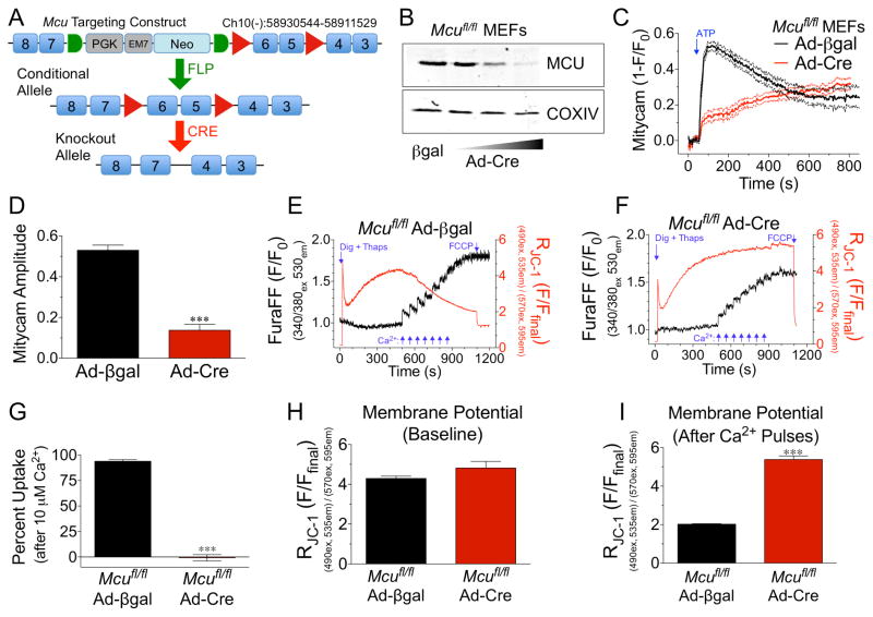
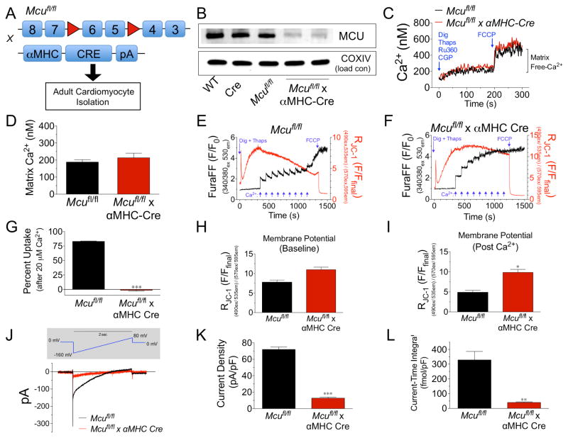
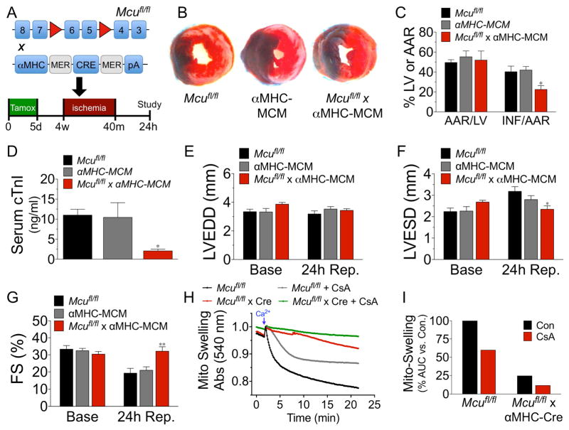
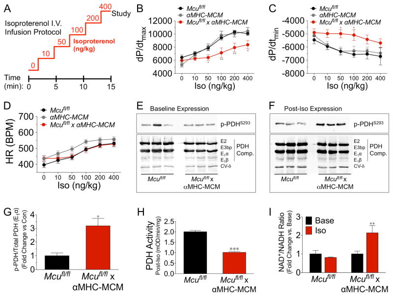
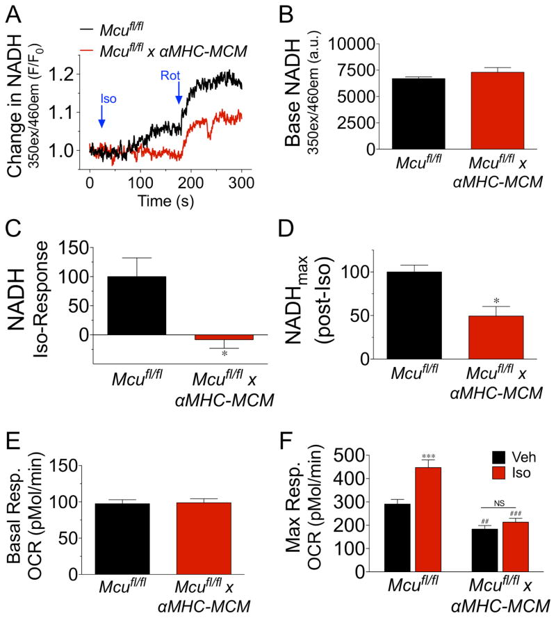
Cardiac contractility is mediated by a variable flux in intracellular calcium (Ca(2+)), thought to be integrated into mitochondria via the mitochondrial calcium uniporter (MCU) channel to match energetic demand. Here, we examine a conditional, cardiomyocyte-specific, mutant mouse lacking Mcu, the pore-forming subunit of the MCU channel, in adulthood. Mcu(-/-) mice display no overt baseline phenotype and are protected against mCa(2+) overload in an in vivo myocardial ischemia-reperfusion injury model by preventing the activation of the mitochondrial permeability transition pore, decreasing infarct size, and preserving cardiac function. In addition, we find that Mcu(-/-) mice lack contractile responsiveness to acute β-adrenergic receptor stimulation and in parallel are unable to activate mitochondrial dehydrogenases and display reduced bioenergetic reserve capacity. These results support the hypothesis that MCU may be dispensable for homeostatic cardiac function but required to modulate Ca(2+)-dependent metabolism during acute stress.


Conflict of interest statement

Conflict of interest: The authors have no financial conflict of interest to report.

Figures

Figure 1
Figure 1. Generation of a conditional Mcu knockout mouse model and confirmation of functionality
A) Schematic of Mcu targeting construct. LoxP sites (red triangles) flank exons 5-6. A neomycin (Neo) selection cassette is flanked by FRT sites (green half-circles). Mutant mice were crossed with ROSA26-FLPe mice for removal of Neo. Floxed mice (conditional allele) were crossed with cardiomyocyte-specific Cre-recombinase driver lines resulting in deletion of Mcu. B) Mouse embryonic fibroblasts (MEFs) were isolated from Mcufl/fl mice at E13.5. MEFs were infected with adenovirus expressing Cre-recombinase (Ad-Cre) or the experimental control β-galactosidase (Ad-βgal). 6d post infection with Ad-Cre cells were lysed and MCU protein expression examined by western blot. COXIV was used as a mitochondrial loading control. C) Mcufl/fl MEFs were treated with Ad-Cre or Ad-βgal and subsequently infected with Adeno encoding mitycam, mCa2+ sensor, 48h prior to imaging. Baseline was recorded and a single pulse of 1-mM ATP was delivered to liberate iCa2+ stores. Signal means shown as solid lines with dashed lines displaying +/− SEM. D) mCa2+ amplitude (peak intensity immediately after ATP – baseline). E) Mcufl/fl MEFs were treated with Ad-βgal and loaded with the Ca2+ sensor (Fura-FF) and the Δψ sensor (JC-1) permeabilized with digitonin and treated with thapsigargin (SERCA inhibitor) for simultaneous ratiometric monitoring during repetitive additions of 5-μM Ca2+ (blue arrows). FCCP was used as a control to collapse Δψ at the conclusion of each experiment. F) Mcufl/fl MEFs were treated with Ad-Cre and subjected to identical experimental conditions. G) Percent mCa2+ uptake vs. Ad-βgal control cells following 10-μM Ca2+ (2nd pulse). H) JC-1 derived Δψ prior to Ca2+ additions. I) JC-1 derived Δψ following 7 pulses of 5-μM Ca2+.
Figure 2
Figure 2. Biophysical characterization of Mcu KO ACMs
A) Mcufl/fl mice were crossed with αMHC-Cre mice and ACMs were isolated from hearts of adult mice. B) ACMs were isolated from: wild-type (WT), αMHC-Cre (Cre), Mcufl/fl, and Mcufl/fl x Cre. Samples were lysed and immunobloted for MCU protein expression and the mitochondrial loading control COXIV. C) ACMs were loaded with the Ca2+ sensor Fura-2. The sarcolemma was permeabilized with digitonin in the presence of thapsigargin (SERCA inhibitor), CGP-37157 (mNCX inhibitor) and Ru360 (MCU inhibitor). Ca2+ levels were recorded and upon reaching a stable baseline, free mCa2+ was released from the mitochondrial matrix with FCCP. D) Quantification of matrix Ca2+ content after Fura calibration. E–F) Mcufl/fl or Mcufl/fl x αMHC-Cre ACMs were loaded with the Ca2+ sensor (Fura-FF) and the Δψ sensor (JC-1) permeabilized with digitonin and treated with thapsigargin (SERCA inhibitor) for simultaneous ratiometric monitoring during repetitive additions of 10-μM Ca2+ (blue arrows). FCCP was used as a control to collapse Δψ at the conclusion of each experiment. G) Percent mCa2+ uptake vs. Mcufl/fl following the addition of 20- μM Ca2+. H–I) JC-1 quantified Δψ at baseline and post-Ca2+ pulses. J) Mitochondria were isolated from ACMs and mitoplasts were prepared for recording of MCU current (iMCU). Voltage ramping protocol (above in grey shaded area) and mean current recordings. K) Current density measured in picoamperes per picofarad (pA/pF). L) Current-time integral measurements, femtomole per picofarad (fmol/pF). (Minimum of 3 independent experiments for all quantified data, all data shown as mean +/− SEM, *p<0.05, ***p<0.001)
Figure 3
Figure 3. Genetic ablation of Mcu protects against myocardial IR-injury
A) Mcufl/fl, αMHC-Mer- Cre-Mer (αMHC MCM) and Mcufl/fl x αMHC-Mer-Cre-Mer mice were treated with tamoxifen (40 mg/kg/day) for 5d to induce cardiomyocyte-restricted Cre expression and allowed to rest for 3wk prior to 40m of ischemia and 24h reperfusion. B) Representative mid-ventricular cross sections of TTC stained hearts. (Evan’s Blue stained area = non-ischemic zone; remaining area = area-at-risk; white area = infracted tissue; red area = viable myocardium.) C) Planimetery analysis of infarct size by quantifying Evan’s blue dye excluded area = area-at-risk (AAR), left ventricle (LV) area and non-TTC stained area = infarct (INF). D) 24h after reperfusion serum was collected and cardiac troponin-I (cTnI) was measured by ELISA. E–G) Mice were analyzed by echocardiography and measurements of LV end-diastolic diameter (LVEDD), LV end-systolic diameter (LVESD), and percent fractional shortening (FS%) were acquired. H) Mitochondria were isolated from hearts of adult mice and changes in swelling (decreased absorbance at 540 nm = increase in volume) were assessed +/− 2-μM CsA. Swelling was initiated by injection of 500-μM Ca2+. I) Changes in swelling quantified by measuring the area-under-the-curve (AUC) and correcting to control. (All in vivo experiments minimum of n=7 for all groups; data shown as mean +/− SEM, *p<0.05, **p<0.01)
Figure 4
Figure 4. mCa2+ uptake is required for β-adrenergic-mediated increases in contractility and bioenergetic responsiveness
A) Mice in all groups received tamoxifen (40 mg/kg/day) for 5d and 1wk later were subjected to an isoproterenol (Iso) infusion protocol (0.1–10 ng/ml) over 15 min. B–D) Invasive hemodynamic analysis of dp/dtmax, dp/dtmin, and heart rate (HR) during Iso infusion (min. n=7/group). E) Baseline expression analysis of pyruvate dehydrogenase (PDH) phosphorylation at S293 of the E1α subunit, and total PDH expression (subunits E2, E3bp, E1α, E1β). ETC Complex V-subunit δ was used as a loading control. F) Hearts were freeze-clamped at the conclusion of Iso infusion protocol and western blot examination of PDH phosphorylation at S293 of the E1α subunit, and total PDH expression (subunits E2, E3bp, E1α, E1β) was performed. G) Fold change in PDH phosphorylation vs. control. Band density analysis calculated as p-PDHS293/total PDH (E1α). H) PDH activity of samples from hearts during Iso administration, expressed as mOD/min/mg of tissue. I) Cardiac NAD+/NADH ratio following Iso infusion, data expressed as fold-change vs. baseline. (All data shown as mean +/− SEM, *p<0.05, **p<0.01, ***p<0.001)
Figure 5
Figure 5. Mcu is necessary for β-adrenergic increases in mitochondrial energetics
ACMs were isolated from Mcufl/fl and Mcufl/fl x αMHC-MCM hearts 1wk post tamoxifen treatment. A) ACMs were monitored spectrofluorometrically for changes in NADH autoflourescence after treatment with isoproterenol (Iso, 10-μM) followed by the addition of rotenone (Rot, 2-μM). Mean NADH recording from 3 independent experiments. B) Baseline NADH levels calculated as fluorescent intensity. C) Percent change in NADH levels following Iso treatment, corrected to Mcufl/fl ACMs. D) NADH fluorescent intensity after treatment with rotenone. Calculated as percent change from baseline to post-rotenone corrected to control ACMs. E) Isolated ACMs were assayed for mitochondrial OxPhos function using a Seahorse Bioanalyzer to measure oxygen consumption rates (OCR). Baseline OCR. F) ACMs were treated with either vehicle (Veh) or isoproterenol (Iso, 10-μM) and FCCP was injected to augment maximal OCR. (All data shown as mean +/− SEM, *p<0.05, ***p<0.001 vs. Mcufl/fl; ##p<0.01, ###p<0.001 vs. Veh)

References

    1. Baines CP, Kaiser RA, Purcell NH, Blair NS, Osinska H, Hambleton MA, Brunskill EW, Sayen MR, Gottlieb RA, Dorn GW, et al. Loss of cyclophilin D reveals a critical role for mitochondrial permeability transition in cell death. Nature. 2005;434:658–662. - PubMed
    1. Baughman JM, Perocchi F, Girgis HS, Plovanich M, Belcher-Timme CA, Sancak Y, Bao XR, Strittmatter L, Goldberger O, Bogorad RL, et al. Integrative genomics identifies MCU as an essential component of the mitochondrial calcium uniporter. Nature. 2011;476:341–345. - PMC - PubMed
    1. Bers DM. Calcium cycling and signaling in cardiac myocytes. Annual review of physiology. 2008;70:23–49. - PubMed
    1. Beutner G, Sharma VK, Giovannucci DR, Yule DI, Sheu SS. Identification of a ryanodine receptor in rat heart mitochondria. The Journal of biological chemistry. 2001;276:21482–21488. - PubMed
    1. Bick AG, Calvo SE, Mootha VK. Evolutionary diversity of the mitochondrial calcium uniporter. Science (New York, N Y. 2012;336:886. - PMC - PubMed

Publication types