

Inflammation Level after Decompression Surgery for a Rat Model of Chronic Severe Spinal Cord Compression and Effects on Ischemia-Reperfusion Injury
- PMID: 26119897
- PMCID: PMC4628191
- DOI: 10.2176/nmc.oa.2015-0022
Inflammation Level after Decompression Surgery for a Rat Model of Chronic Severe Spinal Cord Compression and Effects on Ischemia-Reperfusion Injury
Abstract
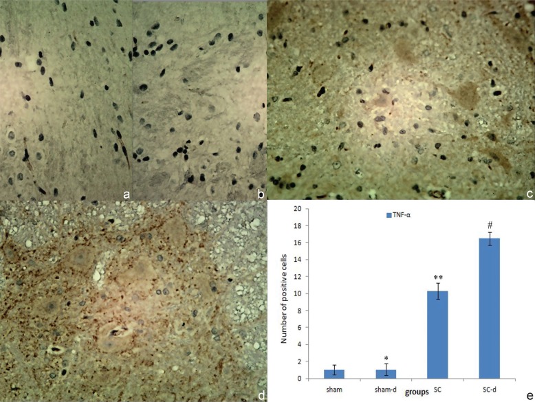
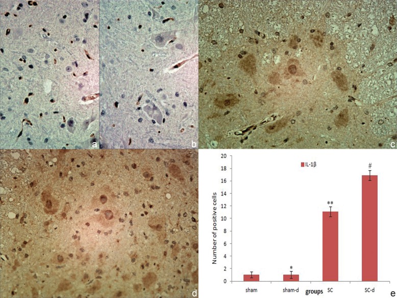
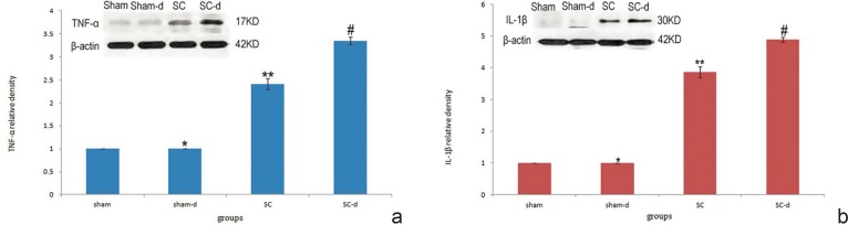
Delayed neurological deterioration in the absence of direct spinal cord insult following surgical decompression is a severe postoperative complication in patients with chronic severe spinal cord compression (SCC). The spinal cord ischemia-reperfusion injury (IRI) has been verified as a potential etiology of the complication. However, the exact pathophysiologic mechanisms of the decompression-related IRI remain to be defined. In this study, we developed a practical rat model of chronic severe SCC. To explore the underlying role of inflammation in decompression-related IRI, immunoreactivity of pro-inflammatory cytokines including tumor necrosis factor-alpha (TNF-α) and interleukin-1β (IL-1β) before and after decompression were measured. In addition, expression level of TNF-α and IL-1β was examined with Western blot. Immunohistochemical staining showed negative result in gray matters in the sham group and sham-decompression group. In the severe compression group, strong positive staining of TNF-α and IL-1β were found, suggesting a dramatic infiltration of inflammatory cells in gray matters. Furthermore, the severe compression group showed a significant increase in expression level of TNF-α and IL-1β as compared with the sham group (p < 0.05). In the severe compression-decompression group, both immunostaining and Western blot showed significant increase of TNF-α and IL-1β levels in the spinal cord compared with the severe compression group (p < 0.05). The results demonstrated that surgical decompression plays a stimulative role in inflammation through increasing the expression of inflammatory cytokines in the rat model of chronic severe SCC injury. Inflammation may be one of the important pathological mechanisms of decompression-related IRI of chronic ischemia.
Conflict of interest statement
The authors report no conflict of interest concerning the materials or methods used in this study or the findings described in this article. No benefits in any form have been or will be received from any commercial party related directly or indirectly to the subject of this manuscript.
Figures
Similar articles
-
Ischemia-reperfusion injury after spinal cord decompressive surgery-An in vivo rat model.Animal Model Exp Med. 2025 Mar;8(3):405-420. doi: 10.1002/ame2.12485. Epub 2024 Sep 3. Animal Model Exp Med. 2025. PMID: 39225110 Free PMC article.
-
Delayed neurological deterioration after surgery for intraspinal meningiomas: Ischemia-reperfusion injury in a rat model.Oncol Lett. 2015 Oct;10(4):2087-2094. doi: 10.3892/ol.2015.3626. Epub 2015 Aug 19. Oncol Lett. 2015. PMID: 26622801 Free PMC article.
-
Anti-inflammatory Mechanism of Bone Marrow Mesenchymal Stem Cell Transplantation in Rat Model of Spinal Cord Injury.Cell Biochem Biophys. 2015 Apr;71(3):1341-7. doi: 10.1007/s12013-014-0354-1. Cell Biochem Biophys. 2015. PMID: 25388837
-
Development and treatments of inflammatory cells and cytokines in spinal cord ischemia-reperfusion injury.Mediators Inflamm. 2013;2013:701970. doi: 10.1155/2013/701970. Epub 2013 Jul 14. Mediators Inflamm. 2013. PMID: 23956505 Free PMC article. Review.
-
Experimental spinal cord trauma: a review of mechanically induced spinal cord injury in rat models.Rev Neurosci. 2017 Jan 1;28(1):15-20. doi: 10.1515/revneuro-2016-0050. Rev Neurosci. 2017. PMID: 27845888 Review.
Cited by
-
Reperfusion injury case following cervical fusion with OPLL: a case report and literature review.J Med Case Rep. 2024 Oct 30;18(1):527. doi: 10.1186/s13256-024-04865-w. J Med Case Rep. 2024. PMID: 39473016 Free PMC article. Review.
-
Brown-Sequard syndrome caused by posterior full-endoscopic cervical discectomy: A case report.Medicine (Baltimore). 2025 Apr 11;104(15):e42007. doi: 10.1097/MD.0000000000042007. Medicine (Baltimore). 2025. PMID: 40228252 Free PMC article.
-
Spinal Cord Parenchyma Vascular Redistribution Underlies Hemodynamic and Neurophysiological Changes at Dynamic Neck Positions in Cervical Spondylotic Myelopathy.Front Neuroanat. 2021 Nov 23;15:729482. doi: 10.3389/fnana.2021.729482. eCollection 2021. Front Neuroanat. 2021. PMID: 34887731 Free PMC article.
-
Letter to the Editor: Commentary on White Cord Syndrome: A Reperfusion Injury Following Spinal Decompression Surgery (Korean J Neurotrauma 2022;18:380-386).Korean J Neurotrauma. 2022 Oct 11;18(2):387-390. doi: 10.13004/kjnt.2022.18.e51. eCollection 2022 Oct. Korean J Neurotrauma. 2022. PMID: 36381442 Free PMC article. No abstract available.
-
White Cord Syndrome Following Long Posterior Decompression.J Orthop Case Rep. 2024 Sep;14(9):14-18. doi: 10.13107/jocr.2024.v14.i09.4712. J Orthop Case Rep. 2024. PMID: 39253650 Free PMC article.
References
-
- Xu P, Gong WM, Li Y, Zhang T, Zhang K, Yin DZ, Jia TH: Destructive pathological changes in the rat spinal cord due to chronic mechanical compression. Laboratory investigation. J Neurosurg Spine 8: 279– 285, 2008 - PubMed
-
- Hasegawa K, Homma T, Chiba Y: Upper extremity palsy following cervical decompression surgery results from a transient spinal cord lesion. Spine (Phila Pa 1976) 32: E197– E202, 2007. - PubMed
-
- Sakaura H, Hosono N, Mukai Y, Ishii T, Yoshikawa H: C5 palsy after decompression surgery for cervical myelopathy: review of the literature. Spine (Phila Pa 1976) 28: 2447– 2451, 2003. - PubMed
-
- Nassr A, Eck JC, Ponnappan RK, Zanoun RR, Donaldson WF, Kang JD: The incidence of C5 palsy after multilevel cervical decompression procedures: a review of 750 consecutive cases. Spine (Phila Pa 1976) 37: 174– 178, 2012. - PubMed
MeSH terms
Substances
LinkOut - more resources
Full Text Sources
Other Literature Sources
Research Materials