Review
. 2015 Jul 27;54(31):8896-927.
doi: 10.1002/anie.201412070. Epub 2015 Jun 26.

Structure-Based Design of Inhibitors of Protein-Protein Interactions: Mimicking Peptide Binding Epitopes

Affiliations
Review

Structure-Based Design of Inhibitors of Protein-Protein Interactions: Mimicking Peptide Binding Epitopes

Marta Pelay-Gimeno et al. Angew Chem Int Ed Engl. .

Abstract

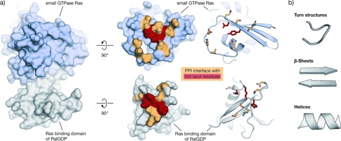
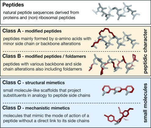
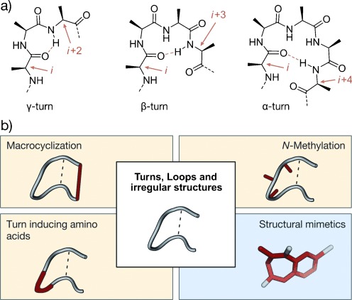
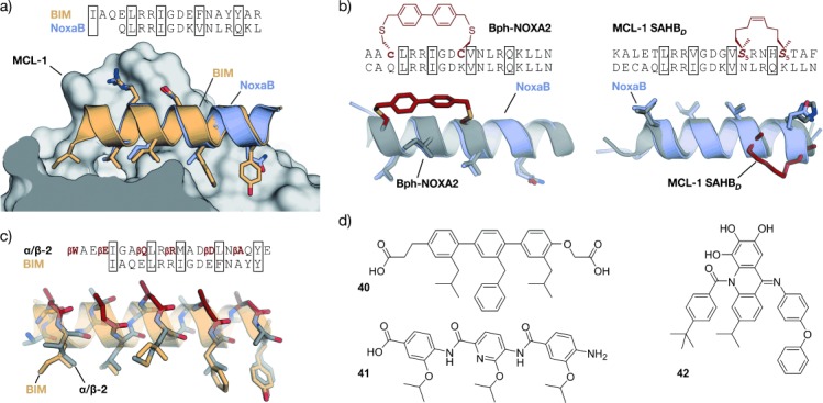
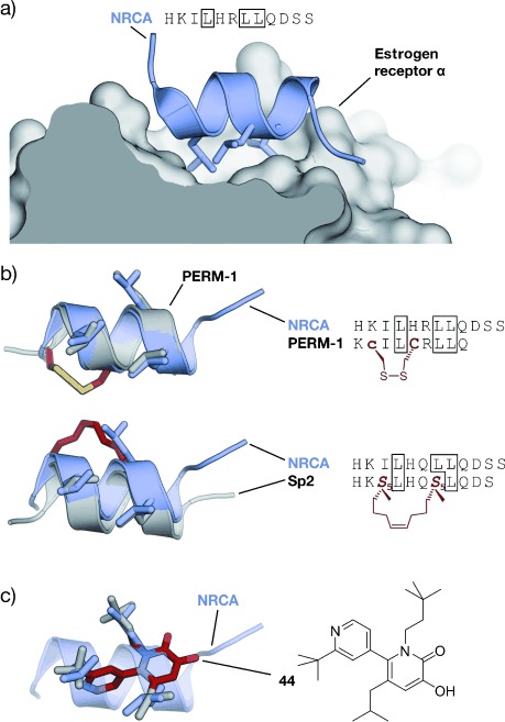
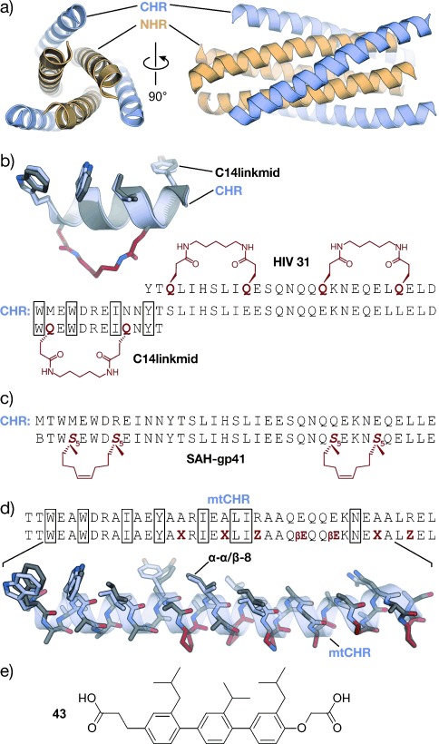
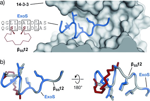
Protein-protein interactions (PPIs) are involved at all levels of cellular organization, thus making the development of PPI inhibitors extremely valuable. The identification of selective inhibitors is challenging because of the shallow and extended nature of PPI interfaces. Inhibitors can be obtained by mimicking peptide binding epitopes in their bioactive conformation. For this purpose, several strategies have been evolved to enable a projection of side chain functionalities in analogy to peptide secondary structures, thereby yielding molecules that are generally referred to as peptidomimetics. Herein, we introduce a new classification of peptidomimetics (classes A-D) that enables a clear assignment of available approaches. Based on this classification, the Review summarizes strategies that have been applied for the structure-based design of PPI inhibitors through stabilizing or mimicking turns, β-sheets, and helices.

Keywords: inhibitors; peptides; peptidomimetics; protein-protein interactions.


Figures

Figure 1
Figure 1
Example of a PPI with its interaction area and hot spots: a) Left: Crystal structure in surface representation of the complex between Ras (light blue) and the Ras binding domain of RalGDP (gray, PDB: 1LFD).[5] Right: The same proteins with their PPI interface (orange/red). Hot-spot residues are highlighted in red. Proteins are shown in surface and schematic representation (interacting residues are shown as sticks);[6] b) Peptide secondary structures in schematic representation.
Figure 2
Figure 2
Classification of peptidomimetics used in this Review: For illustration, an α-helical peptide and corresponding helix mimetics are shown. Modifications are highlighted in red.
Figure 3
Figure 3
Turns with general stabilization and mimicking approaches: a) Chemical structure of a γ-, β-, and α-turn; stabilizing hydrogen bonds are indicated by dotted orange lines, participating residues by orange arrows. b) General strategies for turn stabilization and mimicry (highlighted in red; class A and B: yellow; class C: blue).
Figure 4
Figure 4
Scaffolds that mimic β-turn conformations: Bicyclic peptide (1),[71] benzodiazepine (2),[75] glucose (3),[77] spirocyclic mimetic (4),[82] and trans-pyrollidine-3,4-dicarboxamide (5).[83]
Figure 5
Figure 5
β-Sheets with general stabilization and mimicking approaches: a) Schematic representation of parallel (top) and antiparallel (bottom) β-sheets. b) Chemical structure of a parallel (top) and antiparallel (below) β-sheet arrangement. Hydrogen bonds are represented by dashed lines. c) General strategies to afford β-sheet mimetics (highlighted in red; class A and B: yellow; class C: blue).
Figure 6
Figure 6
β-Sheet mimetics: Turn mimetics: l-Pro-d-Pro (6), dibenzofuran derivatives (7), oligourea (8), azobenzene (9); Macrocyclization: head to tail (10), side chain to side chain cross-link with a disulfide (11) and 1,2,3-triazole ring (12), side chain to side chain π–π interaction (Trp–Trp; 13) and cation–π interaction (Nδ-trimethylornithine–Trp; 14); β-strand-enforcing amino acids: 1,6-dihydro-3(2H)-pyridinone (Ach, 15), Hao building block (16), diphenylacetylene building block (17); Structural mimetics: piperidine–piperidinone-based strand mimetic (18); class A/B: yellow; class C: blue. Box: structure used in the context of PPI inhibition.
Figure 7
Figure 7
Helices with corresponding stabilization and mimicking approaches: a) Chemical structure of a peptide chain, helix-stabilizing hydrogen-bond patterns are indicated by orange arrows. b) Schematic representation of an α-helix together with general strategies of helix stabilization and mimicry (highlighted in red; class A and B: yellow; class C: blue).
Figure 8
Figure 8
Thiol- and lactam-based cross-linked α-helical peptides: a) Disulfide linkage of d- and l-cysteine at positions i, i+3 (19), m-xylene-cross-linked l-cysteines at positions i, i+4 (20), diaryl cross-linked d-cysteine and l-cysteine at positions i, i+7 (21). b) Azobenzene-based cross-linked l-cysteines at positions i, i+7 (22). c) Lactam formed between aspartic acid and lysine at positions i, i+4 (23).
Figure 9
Figure 9
RCM cross-linked α-helical peptides: a) Hydrocarbon-stapled peptides: Cross-linked α-methylated building blocks at positions i, i+4 (24) and i, i+7 (25); b) hydrogen-bond surrogate: Covalent replacement of the hydrogen bond between the N-terminal amino acid (i) and the amine proton at position i+3 (26).
Figure 10
Figure 10
Foldamers: a) Amino acids used in α- and β-peptides as well as peptoids (N-alkylated); b) α- and α/β-peptide in stick representation (schematic representation of the helix is shown transparent). β-Amino acids are highlighted in red; c) β-amino acids commonly used in α/β-peptides (ACPC: trans-2-aminocyclopentanecarboxylic acid, APC: trans-3-aminopyrrolidine-4-carboxylic acid, βD: β3-glutamate as an example of β3-amino acids).
Figure 11
Figure 11
Concept of structural α-helix mimetics (class C): Left: Stick and schematic representations of an α-helix. The side chains i, i+4, and i+7 are represented by a sphere. Right: Stick representation and chemical structure of a terphenyl structural mimetic. The substituents R1, R2, and R3 mimic the tridimensional projection of side chains i, i+4, and i+7 of an α-helix.
Figure 12
Figure 12
Chemical structures of sterically enforced structural α-helix mimetics: A terphenyl (27), two different heterocyclic scaffolds (28 and 29), and a pyrrolopyrimidine (30). The substituents highlighted in gray are designed to mimic the i, i+3/4, and i+7 side chains of an α-helix.
Figure 13
Figure 13
Chemical structures of hydrogen-bond-guided structural α-helix mimetics: An oligopicolinamide or trispyridylamide (31), a 3-O-alkylated oligobenzamide (32), a 2-O-alkylated oligobenzamide (33), an N-alkylated oligobenzamide (34), a terephthalamide (35), and a benzoylurea (36). The substituents highlighted in gray are designed to mimic the i, i+3/4, and i+7 side chains of an α-helix.
Figure 14
Figure 14
Chemical structures of covalently constrained structural α-helix mimetics: An oligooxopiperazine (37) and a spiroligomer (38). The substituents highlighted in gray are designed to mimic the i, i+3/4, and i+7 side chains of an α-helix.
Figure 15
Figure 15
RGD–integrin interaction: a) Crystal structure of the RGD sequence from fibronectin bound to the αV (orange) and β3-subunit (gray) of the integrin receptor (PDB 4MMX). b) Chemical structure of the cyclic pentapeptide cyclo(RGDf-N(Me)V) and crystal structures (gray/red, PDB 1L5G)[341, 342] superimposed with fibronectin RGD (gray; red=constraining amino acids; f=d-phenylalanine).
Figure 16
Figure 16
MDM2–p53 interaction: a) Crystal structures of MDM2 (gray) with the transactivation domain of p53 (blue, PDB 1YCR).[347] b) Superimposed crystal structures of p53 (blue, PDB 1YCR) and cyclic β-hairpin peptide 78A (gray/red, PDB 2AXI). The d-Pro-l-Pro (p-P) cross-link is highlighted in red.[115] c) Sequences of stapled peptides (left). Superimposed crystal structures (right) of p53 (blue, PDB 1YCR) and SAH-p53-8 (gray/red, PDB 3V3B). The cross-link is highlighted in red (side chains of amino acids in boxes are shown explicitly in the crystal structures).[355] d) Superimposed crystal structures of p53 (blue, PDB 1YCR) and Nutlin-3a (red, PDB 4HG7).[356] All superimposed structures were obtained from structures of complexes with MDM2 or MDMX.
Figure 17
Figure 17
PPIs involving proteins of the BCL-2 family: a) Superimposed crystal structures of BIM (orange, PDB 2L9) and NoxaB (blue, PDB 2NLA) bound to MCL-1.[386] b) Superimposed crystal structures of NoxaB (blue, PDB 2NLA) with (left) bisaryl cross-linked peptide Bph-Noxa2 (gray, PDB 4G35, c=d-cysteine)[204] and (right) stapled peptide MCL-1 SAHBD (gray, PDB 3MK8).[387] Cross-links are highlighted in red (side chains of amino acids in boxes are shown explicitly). c) Superimposed crystal structures of BIM (orange, PDB 2L9) and α/β-peptide α/β-2 (gray/red, PDB 4BPI).[388] β-Amino acids are highlighted in red (βE, βQ, βR, βD, and βA are β3-amino acids that correspond to E, Q, R, D, and A, respectively). d) Structural mimetics of helical MCL-1 binding peptides.[312, 389, 390]
Figure 18
Figure 18
Estrogen receptor (ER) coactivator interaction: a) Coactivator peptide NRCA bound to ER α (gray; PDB 2QGT); b) top: superimposed crystal structures of NRCA (blue, PDB 2QGT) and disulfide cross-linked PERM-1 (gray, PDB 1PCG; left). Cys and d-Cys (c) are highlighted in red, the disulfide bridge in yellow; sequences of cross-linked peptide (right). Bottom: Superimposed crystal structures of NRCA (blue, PDB 2QGT) and stapled peptide Sp2 (gray, PDB 2YJA; left). The cross-link is highlighted in red. Sequences of stapled peptide (right). Selected side chains are shown explicitly and highlighted in sequence. c) Superimposed crystal structures of NRCA (blue, PDB 2QGT) and 6-(2-tert-butyl-4-pyridyl)-3-hydroxy-5-isobutyl-1-(3,3-dimethylbutyl)1H-pyridin-2-one (44, gray/red, CCDC: 636896).
Figure 19
Figure 19
Six-helix bundle of gp-41 CHR and NHR helices: a) Crystal structure of the six-helix bundle involving three CHR (blue) and three NHR (orange) helixes (PDB 1AIK).[432] b) Two examples of lactam-bridged stabilized CHR-derived α-helices: HIV 31[434] and C14Linkmid including superimposed crystal structures of a CHR fragment (blue, PDB 1AIK) and C14Linkmid (gray, PDB 1GZL).[216] The amide cross-link is highlighted in red. c) Sequence of CHR-derived double-stapled peptide SAH-gp41.[232] d) Chimeric peptide α-α/β-8 derived from a mutant CHR form (mtCHR). Superimposed crystal structures of mtCHR (blue, PDB 3F4Y) and α-α/β-8 (gray, PDB 3G7A).[439] β-Amino acids are highlighted in red (X=ACPC, Z=APC, βE=β3-glutamate). e) CHR-derived terphenyl structural mimetic[280] (side chains of amino acids in boxes are shown explicitly in the crystal structures).
Figure 20
Figure 20
Interaction between 14-3-3 and ExoS: a) crystal structure of the 14-3-3 binding sequence of exoenzyme S (blue, ExoS) in complex with 14-3-3 (gray, PDB 4N7G). Sequences of ExoS and corresponding cyclic peptide inhibitor βSS12 are shown (side chains of amino acids in boxes are shown explicitly in the crystal structures). b) Overlaid structures of ExoS (blue, PDB 4N7G) and cyclic peptide βSS12 (gray, PDB 4N84). The cross-link is highlighted in red.[92]

References

    1. Milroy LG, Grossmann TN, Hennig S, Brunsveld L, Ottmann C. Chem. Rev. 2014;114:4695–4748. - PubMed
    1. Moreira IS, Fernandes PA, Ramos MJ. Proteins Struct. Funct. Bioinf. 2007;68:803–812. - PubMed
    1. London N, Raveh B, Movshovitz-Attias D, Schueler-Furman O. Proteins Struct. Funct. Bioinf. 2010;78:3140–3149. - PMC - PubMed
    1. Geppert T, Hoy B, Wessler S, Schneider G. Chem. Biol. 2011;18:344–353. - PubMed
    1. Huang L, Hofer F, Martin GS, Kim S-H. Nat. Struct. Mol. Biol. 1998;5:422–426. - PubMed

Publication types

MeSH terms