. 2015 Jul 13;28(1):42-56.
doi: 10.1016/j.ccell.2015.05.007. Epub 2015 Jun 25.

Broad Anti-tumor Activity of a Small Molecule that Selectively Targets the Warburg Effect and Lipogenesis

Affiliations

Broad Anti-tumor Activity of a Small Molecule that Selectively Targets the Warburg Effect and Lipogenesis

Colin A Flaveny et al. Cancer Cell. .

Abstract

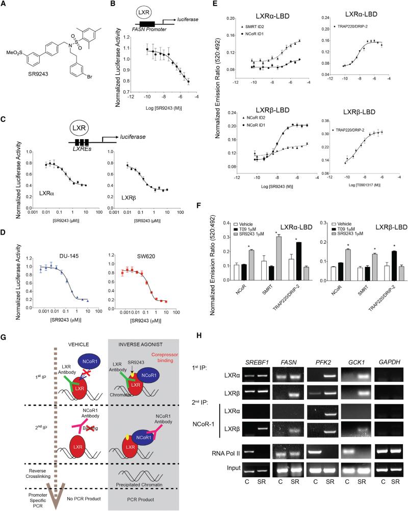
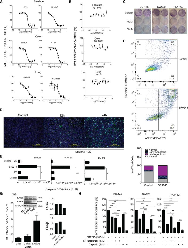
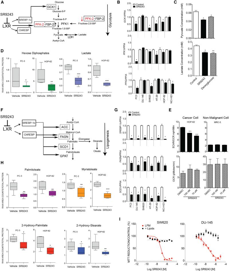
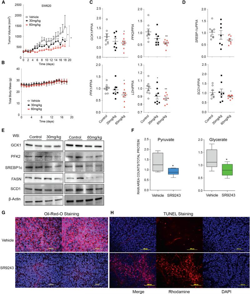
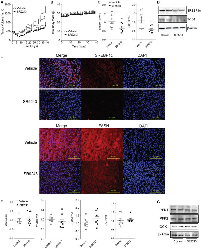
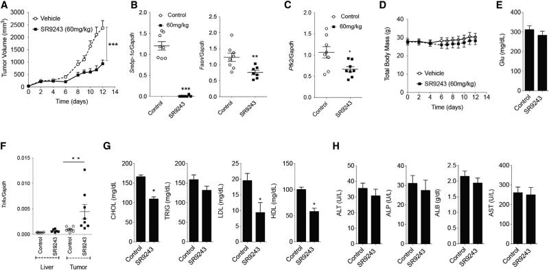
Malignant cells exhibit aerobic glycolysis (the Warburg effect) and become dependent on de novo lipogenesis, which sustains rapid proliferation and resistance to cellular stress. The nuclear receptor liver-X-receptor (LXR) directly regulates expression of key glycolytic and lipogenic genes. To disrupt these oncogenic metabolism pathways, we designed an LXR inverse agonist SR9243 that induces LXR-corepressor interaction. In cancer cells, SR9243 significantly inhibited the Warburg effect and lipogenesis by reducing glycolytic and lipogenic gene expression. SR9243 induced apoptosis in tumors without inducing weight loss, hepatotoxicity, or inflammation. Our results suggest that LXR inverse agonists may be an effective cancer treatment approach.


Figures

Figure 1
Figure 1. SR9243 Is an LXR Inverse Agonist that Induces Corepressor Recruitment
(A) Structure of SR9243. (B) FASN promoter-driven luciferase reporter assay showing SR9243 repression of the basal activity of exogenously expressed full-length LXRα or LXRβ in HEK293 cells. Transfected cells were treated with 0.01–10 μM of SR9243 for 6 hr. (C) LXRE-driven (3X) luciferase reporter assay showing SR9243 repression of basal activity of endogenously expressed LXRs in HEK293 cells. Cells were treated with 1 nM–10 μM SR9243 for 6 hr. (D) LXRE-driven luciferase reporter dose-response assays in cancer cell lines expressing exogenous LXRα or LXRβ and treated with SR9243. (E) TR-FRET assay showing the recruitment of corepressor box peptides NCoR and SMRT to LXRα and LXRβ in response to SR9243. (F) TR-FRET assay showing relative recruitment of NCoR, SMRT, and TRAP220/DRIP-2 box peptides to LXRα or LXRβ LBDs in response to vehicle of SR9243 or 1 μM of the LXR agonist T0901317. (G) Diagram illustrating the Re-ChIP experiment shown in (H) PCR results of Re-ChIP assays of LXRα or LXRβ followed by NCOR1 showing SR9243 (SR)-induced corepressor recruitment at the promoters of GCK1, PFK2, SREBF, and FASN. (C, control; SR, SR9243) Representative figure of experiment repeated three times. *p < 0.05, **p < 0.01, ***p < 0.001. Error bars represent ±SEM. See also Figure S1.
Figure 2
Figure 2. SR9243 Induces Apoptotic Cell Death in Multiple Cancer Cell Types in an LXR-Dependent Manner
(A) MTT assay showing the viability of cancer cells from the prostate (PC3, DU-145), colon (SW620, HT-29), and lung (HOP-62, NCI-H23) treated with increasing doses of SR9243. (B) MTT assay of non-malignant cells from the prostate (PZ-HPV-7), colon (CCD-18Co), and lung (MRC5) treated with SR9243. (C) Colony formation assay of cancer cells treated with vehicle, or 100 nM or 10 μM SR9243. (D) Fluorescent cell viability/apoptosis assay showing the viable cells (blue) and or the apoptotic cells (green) from SW620 cells treated with 1 μM SR9243 for 12 and 24 hr. (E) Luminescence-based caspase 3/7 activity of SW620, HOP62, and DU-145 cells treated with vehicle, 1 μM SR9243, and or the caspase inhibitor (Z-VAD-FMK) for 24 hr. (F) FACs sorting of Annexin V FITC/PI-stained SW620 cells treated with SR9243 (100 nM) for 24 hr. The percentage of apoptotic, necrotic and live cell populations are graphically represented in lower image. (G) MTT reduction assay showing rescue of cell viability of SW620 colon cancer cells transfected with LXRα and LXRβ-specific siRNAs compared to control or mock transfected cells. LXR expression in SW620 cells treated with LXR siRNAs (right). (H) MTT reduction assay showing the viability of DU-145, SW620, and HOP-62 cancer cells in response to SR9243 alone or in combination with 5′-fluorouracil or cisplatin. Experiments were repeated three times. *p < 0.05, **p < 0.01, ***p < 0.001. Error bars represent ±SEM. See also Figure S2.
Figure 3
Figure 3. SR9243 Inhibits Glycolytic Lipogenic Enzymes in Cancer Cells
(A) Schematic showing glycolysis genes regulated by LXRs and associated metabolites. Gene names outlined in boxes are directly regulated by LXRs. (B) RT-PCR showing expression of LXR-regulated glycolysis genes GCK1, PFK1, and PFK2 after 6 hr treatment with SR9243 (10 μM). (C) Biochemically determined cellular levels of lactate and pyruvate in HOP-62 cancer cells treated with SR9243 for 24 hr. (D) GC/MS determined levels of lactate and hexose phosphates in PC3 and HOP62 cancer cells. (E) Relative glycolytic rate (ECAR/OCR) and oxygen consumption rate (OCR) of HOP-62 lung cancer cells or the corresponding non-tumorigenic lung epithelial cells (MRC5) treated with SR9243 (100 nM or 10 μM). Cells were assessed for extracellular acidification rate and OCR using the Seahorse XF extracellular flux analyzer. (F) Schematic showing the lipogenesis genes regulated by LXR and their cognate substrates and products. RT-PCR showing expression profile of LXR-regulated lipogenesis genes SREBP1c, FASN, and SCD1 in cancer cells treated with SR9243 (10 μM) for 6 hr. (G and H) (G) GC/MS showing cellular levels of the long-chain fatty acids palmitoleate and myristoleate in SR9243 treated cancer cells and (H) the short-chain fatty acids (2-hydroxy-palmitate and 2-hydroxy-stearate) in SW620 and HOP-62 cells treated with SR9243. Samples were normalized by total protein concentration. (I) MTT assay of DU-145 and SW620 cells treated with increasing amounts of SR9243 in LFM (lipid-free media) or positive lipid media (supplemented with 25 nM oleate, palmitate, and stearate) for 96 hr. Data were analyzed using Welch's t tests and/or Wilcoxon's rank sum tests. *p < 0.05, *p < 0.05, **p < 0.01, ***p < 0.001. Error bars represent ±SEM. See also Figure S3.
Figure 4
Figure 4. SR9243 Reduces Tumor Growth, Glycolytic and Lipogenic Enzyme Expression in Tumors In Vivo
(A) Volume of SW620 colon cancer xenografts in athymic mice treated with vehicle (n = 9), 30 mg/kg (n = 9), or 60 mg/kg (n = 9) SR9243. (B) Total body weight of mice from (A). (C and D) RT-PCR analysis of the (C) glycolytic genes GCK1, PFK1, and PFK2 and (D) the lipogenesis genes SREBP1c and SCD1 in tumors of mice treated with 30 and 60 mg/kg SR9243. RT-PCR data were analyzed using Student's t test. *p < 0.05. (E) Immunoblot showing expression levels of SREBP1c, FASN, SCD1, GCK1, and PFK2 protein in SW620 tumors treated with 30 or 60 mg/kg SR9243. (F) GC/MS determined concentration of pyruvate and glycerate in SW620 xenografts in mice treated with 30 mg/kg SR9243. Data were analyzed using Welch's t tests and/or Wilcoxon's rank sum tests. *p < 0.05. Error bars represent ±SEM. (G) Oil red O staining showing lipid content of SW620 tumors in mice treated with 60 mg/kg SR9243 or vehicle. (H) Fluorescence based in situ TUNEL assay showing apoptotic cells in tumors from mice treated with SR9243 (60 mg/kg) or vehicle control. See also Figure S4.
Figure 5
Figure 5. SR9243 Treatment Inhibits Lipogenic Enzyme Expression to Suppress Tumor Growth
(A) Volume of prostate cancer (DU-145)-xenografts in nude mice treated with 60 mg/kg SR9243 (n = 8) or vehicle (n = 7). (B) Total body weight of mice treated in (A). (C) RT-PCR showing expression of SREBP1c and SCD1 in tumors from mice treated with SR9243 or vehicle. Data were analyzed using Student's t test followed by Bonferroni post-test. *p < 0.05. Error bars represent ±SEM). (D) Immunoblot showing protein expression of SREBP1c and SCD1 in control and SR9243-treated tumors. (E) Fluorescence-based immunohistochemistry SREBP1c and FASN expression in tumors from vehicle and SR9243-treated mice. (F) RT-PCR showing expression of PFK1, PFK2, GCK1, and LDH in tumors treated with SR9243 or control. Data were analyzed using Student's t test followed by Bonferroni post-test. *p < 0.05. (G) Immunoblot showing protein expression of SREBP1c and SCD1 in control and SR9243-treated tumors and SCD1 in tumors from mice treated with SR9243 or vehicle. See also Figure S5.
Figure 6
Figure 6. SR9243 Inhibits Tumor Growth and Lipogenesis without Hepatotoxicity or Inflammation
(A) Volume of mouse Lewis lung carcinoma (LLC1) syngeneic tumors implanted in C57BL6/J mice treated with vehicle (n = 8) or 60 mg/kg SR9243 (n = 8). (B and C) RT-PCR analysis of (B) Srebp1c, Fasn, and (C) Pfk2 expression in tumors from mice treated with SR9243 or vehicle. (D) Total body weight of tumor-bearing mice treated with SR9243. (E) Serological analysis of blood glucose levels in vehicle and SR9243-treated mice. (F) RT-PCR analysis showing expression of the cytokine Tnfα in livers and LLC1 tumors of C57BL6J mice treated with SR9243. See also Figure S5. (G) Plasma levels of total cholesterol (CHOL), triglycerides (TRIG), low-density lipoproteins (LDL), and high-density lipoproteins (HDL) in vehicle and SR9243 (60 mg//kg)-treated mice. (H) Plasma levels of the liver transaminases aspartate transaminase (AST), alkaline phosphatase (ALP), and alanine aminotransferase (ALT) as well as total albumin (ALB) in vehicle and SR9243-treated tumor-bearing mice. All RT-PCR and serological data were analyzed using a two-tailed t test followed by Bonferroni post-test. *p < 0.05. Error bars represent ±SEM. See also Figure S6.

Comment in

References

    1. Baranowski M. Biological role of liver X receptors. J. Physiol. Pharmacol. 2008;59(Suppl 7):31–55. - PubMed
    1. Beyea MM, Heslop CL, Sawyez CG, Edwards JY, Markle JG, Hegele RA, Huff MW. Selective up-regulation of LXR-regulated genes ABCA1, ABCG1, and APOE in macrophages through increased endogenous synthesis of 24(S),25-epoxycholesterol. J. Biol. Chem. 2007;282:5207–5216. - PubMed
    1. Blancher C, Harris AL. The molecular basis of the hypoxia response pathway: tumour hypoxia as a therapy target. Cancer Metastasis Rev. 1998;17:187–194. - PubMed
    1. Bonuccelli G, Tsirigos A, Whitaker-Menezes D, Pavlides S, Pestell RG, Chiavarina B, Frank PG, Flomenberg N, Howell A, Martinez-Outschoorn UE, et al. Ketones and lactate “fuel” tumor growth and metastasis: Evidence that epithelial cancer cells use oxidative mitochondrial metabolism. Cell Cycle. 2010;9:3506–3514. - PMC - PubMed
    1. Carracedo A, Cantley LC, Pandolfi PP. Cancer metabolism: fatty acid oxidation in the limelight. Nat. Rev. Cancer. 2013;13:227–232. - PMC - PubMed

MeSH terms