. 2015 Jun 12:21:673-87.
eCollection 2015.

Characterization of cells from patient-derived fibrovascular membranes in proliferative diabetic retinopathy

Affiliations

Characterization of cells from patient-derived fibrovascular membranes in proliferative diabetic retinopathy

Leo A Kim et al. Mol Vis. .

Abstract

Purpose: Epiretinal fibrovascular membranes (FVMs) are a hallmark of proliferative diabetic retinopathy (PDR). Surgical removal of FVMs is often indicated to treat tractional retinal detachment. This potentially informative pathological tissue is usually disposed of after surgery without further examination. We developed a method for isolating and characterizing cells derived from FVMs and correlated their expression of specific markers in culture with that in tissue.

Methods: FVMs were obtained from 11 patients with PDR during diabetic vitrectomy surgery and were analyzed with electron microscopy (EM), comparative genomic hybridization (CGH), immunohistochemistry, and/or digested with collagenase II for cell isolation and culture. Antibody arrays and enzyme-linked immunosorbent assay (ELISA) were used to profile secreted angiogenesis-related proteins in cell culture supernatants.

Results: EM analysis of the FVMs showed abnormal vessels composed of endothelial cells with large nuclei and plasma membrane infoldings, loosely attached perivascular cells, and stromal cells. The cellular constituents of the FVMs lacked major chromosomal aberrations as shown with CGH. Cells derived from FVMs (C-FVMs) could be isolated and maintained in culture. The C-FVMs retained the expression of markers of cell identity in primary culture, which define specific cell populations including CD31-positive, alpha-smooth muscle actin-positive (SMA), and glial fibrillary acidic protein-positive (GFAP) cells. In primary culture, secretion of angiopoietin-1 and thrombospondin-1 was significantly decreased in culture conditions that resemble a diabetic environment in SMA-positive C-FVMs compared to human retinal pericytes derived from a non-diabetic donor.

Conclusions: C-FVMs obtained from individuals with PDR can be isolated, cultured, and profiled in vitro and may constitute a unique resource for the discovery of cell signaling mechanisms underlying PDR that extends beyond current animal and cell culture models.


Figures

Figure 1
Figure 1
Clinical case 3 representing combined tractional and rhegmatogenous retinal detachment with resection of FVM and growth of C-FVM. Pre-operative fundus photograph of the left eye reveals combined tractional and rhegmatogenous retinal detachment of the left eye with 20/200 vision (A). FVM (outlined by dotted white line) involves the macula and reveals fibrous tissue with aberrant blood vessels. Post-operative fundus photograph five months post vitrectomy and one month after cataract surgery demonstrates re-attached retina with 20/25 vision (B). Bright field microscopy of surgically resected FVM shows fibrous tissues and blood vessels (C, inset). Bright field microscopy of cells derived from FVM reveals a morphologically heterogeneous mixture of fibroblastic and stellate cells (D). Scale bar = 100 µm.
Figure 2
Figure 2
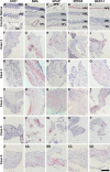
Histopathological characterization of FVM tissue from patients with severe PDR. Light micrographs of the control retina (A–E) and fibrovascular membranes (FVMs) from five different cases (F–DD) processed for immunohistochemistry using primary antibodies against CD 31 (A, F, K, P, U, Z), smooth muscle actin (SMA, B, G, L, Q, V, AA), glial fibrillary acidic protein (GFAP, C, H, M, R, W, BB), retinal pigment epithelium-specific protein 65 (RPE65, D, I, N, S, X, CC), and bestrophin-1 (BEST-1, E, J, O, T, Y, DD; all in red) and counterstained with hematoxylin (blue). In the control retina, faint CD31 staining of endothelial cells is observed (A), SMA localization is restricted to vessels (B), GFAP staining localizes to cells surrounding the blood vessels within the nerve fiber layer (NFL, C), and RPE65 and BEST-1 localize to the RPE cells (D, E). For FVMs derived from different patients, however, there was notable variability in the localization patterns and the number of positive cells for the different markers. All FVMs have CD31-positive cells, which are perivascular (F, K, P, U, Z). SMA-positive cells were also detected in all FVMs; some SMA staining was localized predominately to cells surrounding the vessels (G, AA) whereas other SMA-positive cells appear to be stromal (L, Q, V), located throughout the fibrous tissue of the FVM. GFAP-positive cells are also observed in all FVMs; some GFAP-positive cells are seen surrounding vessels (H, BB) whereas other GFAP-positive cells are located within the stroma of the FVM (M, R, W). Whereas all FVMs are RPE65-negative (I, N, S, X, CC), BEST-1 positive cells are distributed throughout the FVM tissue, as well as localized to perivascular cells (J, O, T, Y, DD). Scale bar=100 µm.
Figure 3
Figure 3
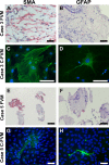
Histopathological characteristics of FVMs are retained in C-FVMs. Fibrovascular membranes (FVMs) from clinical cases 3 and 5 were processed for immunohistochemistry using antibodies against SMA (A, E) and GFAP (B, F; all in red) and counterstained with hematoxylin (blue). Immunofluorescence of the C-FVMs shows cells positive for SMA (C, G) and GFAP (D, H; in green), indicating that molecular markers of cell identity are retained in C-FVMs grown in vitro. Nuclei are counterstained with 4',6-diamidino-2-phenylindole (DAPI; blue). Scale bar=50 µm.
Figure 4
Figure 4
Proliferative activity of C-FVMs varies between different clinical cases. Fibrovascular membranes (FVMs) from clinical cases 3 (A) and 5 (B) were processed for immunohistochemistry using antibodies against Ki67 (in red) and counterstained with hematoxylin (blue). C-FVMs from clinical case 3 and clinical case 5 differ in the percentage of cells that are Ki67-negative (66.06% versus 81.60%) and Ki67-positive (33.94% versus 18.40%) when cultured in growth media (C, D). The percentage of cells that are Ki67-negative (92.57% versus 94.06%) and Ki67-positive (7.43% versus 5.94%) in C-FVMs from clinical cases 3 and 5 are similar under growth factor-starvation conditions (E, F). Scale bar=50 µm.
Figure 5
Figure 5
Isolation of CD31-positive cells from C-FVMs. Immunofluorescence localization of isolectin (A) and CD31 (B) cells from clinical case 4 identifies rare positive cells. Fibrovascular membrane (FVM) from clinical case 5 processed for immunohistochemistry using antibodies against CD31 (red) and counterstained with hematoxylin (blue; C). Light micrographs of C-FVMs from clinical case 5 show isolation of CD31-positive cells, bound to CD31-antibody coated Dynabeads before cell culture (D). Scale bar=50 µm.
Figure 6
Figure 6
Angiogenesis-related proteins secreted by C-FVMs: The effect of high glucose and hyperosmolarity. Map of the human angiogenesis antibody array targeting 55 proteins related to angiogenesis and angiogenesis-related factors identified 11 proteins secreted by C-FVMs from clinical case 3 (A). For identity and coordinates of all 55 proteins assayed, see Appendix 4. Protein levels of (B) angiopoietin-1, (C) thrombospondin-1, (D) PEDF, and (E) endothelin-1 under different glucose treatments were measured in human retinal pericytes (HRP) and C-FVMs from clinical case 3 using an ELISA assay. Student t tests show there was a significant reduction in levels of angiopoietin-1 and thrombospondin-1 by cells grown in high glucose (30 mM D-glucose) and under hyperosmolar conditions (30 mM L-glucose), as compared to normal glucose and normal osmolar conditions (5 mM). No changes in PEDF or endothelin-1 protein levels were observed in C-FVMs in response to changes in glucose or osmolarity conditions. The levels of angiopoietin-1, thrombospondin-1, PEDF, and endothelin-1 did not in change in HRP in response to treatment with different glucose or osmolar conditions. Error bars represent ± standard deviations; **p<0.01; n=3.

References

    1. Cheung N, Mitchell P, Wong TY. Diabetic retinopathy. Lancet. 2010;376:124–36. - PubMed
    1. Klein R, Klein BE, Moss SE, Davis MD, DeMets DL. The Wisconsin epidemiologic study of diabetic retinopathy. III. Prevalence and risk of diabetic retinopathy when age at diagnosis is 30 or more years. Arch Ophthalmol. 1984;102:527–32. - PubMed
    1. Yau JW, Rogers SL, Kawasaki R, Lamoureux EL, Kowalski JW, Bek T, Chen SJ, Dekker JM, Fletcher A, Grauslund J, Haffner S, Hamman RF, Ikram MK, Kayama T, Klein BE, Klein R, Krishnaiah S, Mayurasakorn K, O'Hare JP, Orchard TJ, Porta M, Rema M, Roy MS, Sharma T, Shaw J, Taylor H, Tielsch JM, Varma R, Wang JJ, Wang N, West S, Xu L, Yasuda M, Zhang X, Mitchell P, Wong TY. Global prevalence and major risk factors of diabetic retinopathy. Diabetes Care. 2012;35:556–64. - PMC - PubMed
    1. Antonetti DA, Klein R, Gardner TW. Diabetic retinopathy. N Engl J Med. 2012;366:1227–39. - PubMed
    1. Eliott D, Lee M, Abrams GW. Proliferative diabetic retinopathy: principles and techniques of surgery. In: Ryan SJ, editor. Retina. 4th ed: Elsevier; 2005.

Publication types

MeSH terms